Chapter: Human Nervous System and Sensory Organs : Brain Stem and Cranial Nerves
Eye-Muscle Nerves (Cranial Nerves III, IV, and VI)

Eye-Muscle Nerves (Cranial Nerves III, IV, and VI)
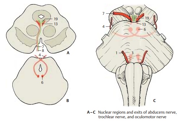
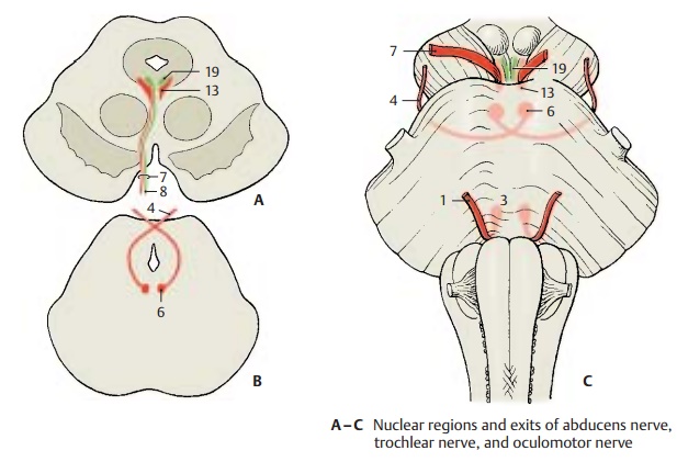
Abducens Nerve (C, E)
The sixth cranial nerve (C1) is an exclu-sively somatomotor nerve, which innervates the lateral rectus muscle (E2) of the extra-ocu-lar muscles. Its fibers originate from thelarge, multipolar neurons of the nucleus ofthe abducens nerve (C3), which lies in thepons in the floor of the rhomboid fossa. The fibers exit at the basal mar-gin of the pons above the pyramid. After taking a long intradural course, the nerve passes through the cavernous sinus and leaves the cranial cavity through the supe-rior orbital fissure.

Trochlear Nerve (B, C, E)
The fourth cranial nerve (BC4) is an exclu-sively somatomotor nerve and innervates the superior oblique muscle (E5) of the extra-ocular muscles. Its fibers originate from thelarge, multipolar neurons of the nucleus ofthe trochlear nerve (BC6), whichlies in the midbrain below the aqueduct at the level of the inferior colliculi. The fibers ascend dorsally in an arch, cross above the aqueduct, and leave the midbrain at the lower margin of the inferior colliculi. The nerve is the only cranial nerve leaving the brain stem at its dorsal aspect. It descends in the subarachnoid space to the base of the skull, where it enters the dura mater at the margin of the tentorium and continues through the lateral wall of the cavernous sinus. It enters the orbit through the superior orbital fissure.

Oculomotor Nerve (A, C – F)
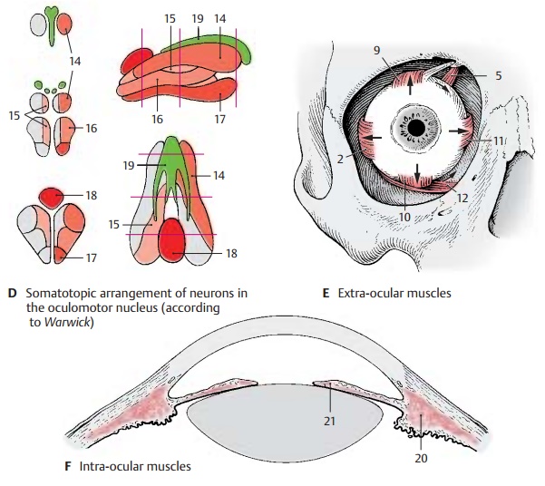
The third cranial nerve (AC7) contains so-matomotor and visceromotor (parasympa-thetic) (A8) fibers. It innervates the remain-ing outer eye muscles (E) and, with itsvisceromotor portion, the intra-ocularmuscles. The fibers exit from the floor of theinterpeduncular fossa at the medial margin of the cerebral peduncle in the oculomotorsulcus. Laterally to the sella turcica, they penetrate the dura mater, run through the roof and then through the lateral wall of the cavernous sinus, and enter the orbit through the superior orbital fissure. Here, the nerve divides into a superior branch, which supplies the levator muscle of theupper eyelid and the superior rector muscle (E9), and into an inferior branch, which sup-plies the inferior rector muscle (E10), the me-dial rector muscle (E11), and the inferior ob-lique muscle (E12).The somatomotor fibers originate from large multipolar neurons of the nucleus of theoculomotor nerve (AC13), whichlies in the midbrain below the aqueduct at the level of the superior colliculi.
The longitudinally arranged cell groups in-nervate specific muscles. The neurons for the inferior rector muscle (D14) lie dor-solaterally, those for the superior rector muscle (D15) dorsomedially; below them lie the neurons for the inferior oblique muscle (D16), those for the medial rector muscle (D17) ventrally, and those for the le-vator muscle of the upper eyelids (caudal central oculomotor nucleus) (D18) dorso-caudally. In the middle third between the two paired main nuclei there usually lies an unpaired cell group, Perlia’s nucleus, which is thought to be associated with ocular con-vergence.
The preganglionic visceromotor (parasym-pathetic) fibers originate from the parvo-cellular Edinger – Westphal nucleus, the acces-sory oculomotor nucleus (ACD19). They runfrom the oculomotor nucleus to the ciliary ganglion where they synapse. The postgan-glionic fibers enter through the sclera into the eyeball and innervate the ciliary muscle (F20) and the sphincter pupillae muscle(F21).
Related Topics