Chapter: Medical Surgical Nursing: Management of Patients With Dysrhythmias and Conduction Problems
Dysrhythmias: Normal Electrical Conduction

Dysrhythmias
Dysrhythmias are disorders of the
formation or conduction (or both) of the electrical impulse within the heart.
These disorders can cause disturbances of the heart rate, the heart rhythm, or
both. Dysrhythmias may initially be evidenced by the hemodynamic effect they
cause (eg, a change in conduction may change the pumping action of the heart
and cause decreased blood pressure).Dysrhythmias are diagnosed by analyzing the
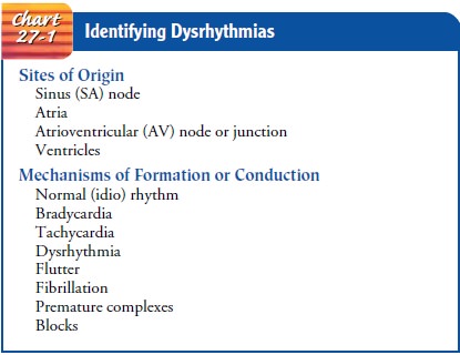
electrocardiographic waveform. They are named according to the site of origin
of the impulse and the mechanism of formation or conduction involved (Chart
27-1). For example, an impulse that originates in the sinoatrial (SA) node and
that has a slow rate is called sinus bradycardia.

NORMAL ELECTRICAL CONDUCTION
The electrical impulse that stimulates
and paces the cardiac muscle normally originates in the sinus node (SA node),
an area located near the superior vena cava in the right atrium. Usually, the
elecrical impulse occurs at a rate ranging between 60 and 100 times a minute in
the adult. The electrical impulse quickly travels from the sinus node through
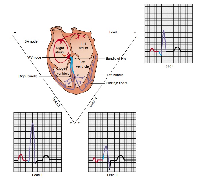
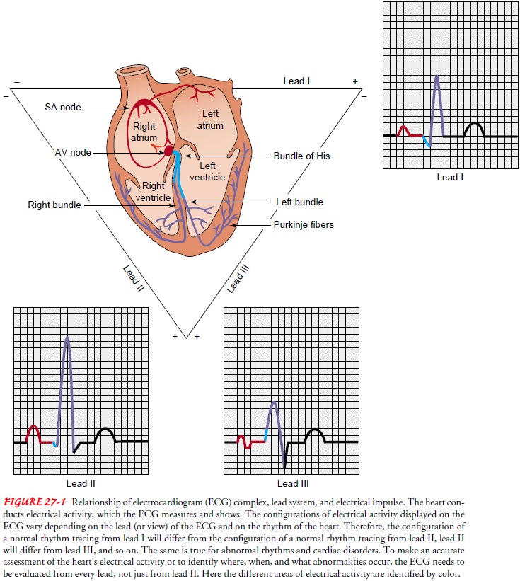
the atria to the atrioventricular (AV) node (Fig. 27-1). The electrical
stimulation of the muscle cells of the atria causes them to contract. The
structure of the AV node slows the electrical impulse, which allows time for
the atria to contract and fill the ventricles with blood before the electrical
impulsetravels very quickly through the bundle of His to the right and left
bundle branches and the Purkinje fibers, located in the ventricular muscle. The
electrical stimulation of the muscle cells of the ventricles, in turn, causes
the mechanical contraction of the ventricles (systole). The cells repolarize
and the ventricles then relax (diastole). The process from sinus node
electrical impulse generation through ventricular repolarization completes the
electro mechanical circuit, and the cycle begins again.
Sinus rhythm promotes cardiovascular
circulation. The electrical impulse causes (and, therefore, is followed by) the
mechanical contraction of the heart muscle. The electrical stimulation is
called depolarization; the mechanical contraction is called systole. Electrical
relaxation is called repolarization and mechanical relaxation is called
diastole.

Influences on Heart Rate and Contractility
The
heart rate is influenced by the autonomic nervous system, which consists of sympathetic
and parasympathetic fibers. Sym-pathetic nerve fibers (also referred to as
adrenergic fibers) are at-tached to the heart and arteries as well as several
other areas in the body. Stimulation of the sympathetic system increases heart
rate (positive chronotropy), conduction through the AV node (positive
dromotropy), and the force of myocardial contraction (positive inotropy).
Sympathetic stimulation also constricts peripheral blood vessels, therefore
increasing blood pressure. Parasympathetic nerve fibers are also attached to
the heart and arteries. Parasympathetic stimulation reduces the heart rate
(neg-ative chronotropy), AV conduction (negative dromotropy), and the force of
atrial myocardial contraction. The decreased sym-pathetic stimulation results
in dilation of arteries, thereby low-ering blood pressure.
Manipulation
of the autonomic nervous system may increase or decrease the incidence of
dysrhythmias. Increased sympathetic stimulation—caused, for example, by
exercise, anxiety, fever, or administration of catecholamines (eg, dopamine
[Intropin], aminophylline, dobutamine [Dobutrex])—may increase the in-cidence
of dysrhythmias. Decreased sympathetic stimulation (eg, with rest,
anxiety-reduction methods such as therapeutic com-munication or prayer,
administration of beta-adrenergic block-ing agents) may decrease the incidence
of dysrhythmias.
Related Topics