Chapter: Human Nervous System and Sensory Organs : Brain Stem and Cranial Nerves
Cross Section Through the Superior Colliculi of the Midbrain

Cross Section Through the Superior Colliculi of the Midbrain
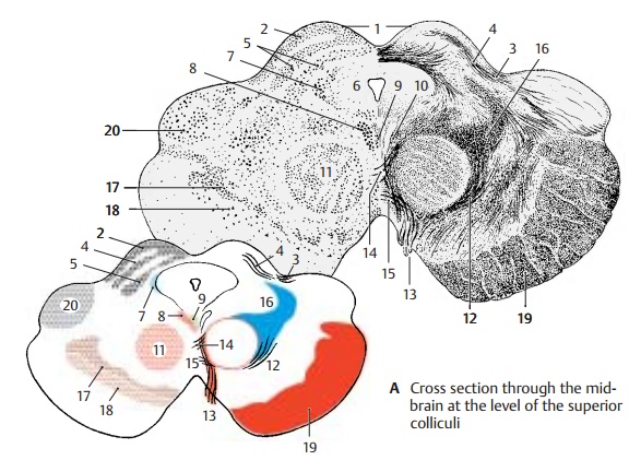
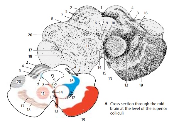
The two superior colliculi (A1) are seen dorsally. In lower vertebrates, they represent the
most important visual center and consist of several layers of cells and
fibers. In humans, they are only a relay station for re-flex movements of the
eyes and pupillary reflexes, and their stratification is rudimen-tary. In the superficial gray layer (A2) termi-nate the fibers from the
occipital fields of the cortex (corticotectal
tract) (A3). The optic layer (A4), which in lower vertebratesconsists of fibers of the optical
tract, is formed in humans by fibers from the lateral genicular body. The
deeper layers of cells and fibers are collectively known as stratumlemnisci (A5). Here terminate the spinotec-tal tract, fibers of the medial and lateral
lemnisci, and fiber bundles of the in-ferior colliculi.

The aqueduct is surrounded by the
peri-aqueductal gray, or central gray
(AB6). It contains a large number of
peptidergic neu-rons (VIP, enkephalin, cholecystokinin, and others). The mesencephalic nucleus of thetrigeminal
nerve (A7) lies laterally to it,
andventrally to it lie the nucleus of
the oculomotornerve (A8) and the Edinger-Westphal nucleus (accessory
oculomotor nucleus) (A9). Dorsally
to both nuclei runstheposterior longitudinalfasc iculus(Schütz’s bundle) and ventrally to them the medial longitudinal fasciculus(A10). The main nucleus of the
teg-mentum is the red nucleus (AB11); it is delimited by a capsule
con-sisting of afferent and efferent fibers (among others, the dentatorubral fasciculus) (A12). At its medial margin descend fiberbundles of the oculomotor nerve (A13) inventral direction. Tectospinal
fibers (pupil-lary reflex) and tectorubral fibers cross the midline in the superior tegmental decussa-tion (Meynert’s decussation) (A14) and teg-mentospinal fibers in the inferior decussa-tion (Forel’s decussation) (A15). The lateralfield is occupied by
the medial lemniscus (AB16).
Ventrally to the tegmentum border
the sub-stantia nigra (pars compacta[A17] andparsreticulata [A18], p.136; p. 148, A1). The ven-tral
aspect on both sides is formed by the corticofugal fiber masses of the cerebralpeduncles (AB19). The dorsal aspect isformed by the medial genicular body (AB20).
Related Topics