Chapter: Basic Concept of Biotechnology : Animal Biotechnology
Cell Cycle - Animal Biotechnology
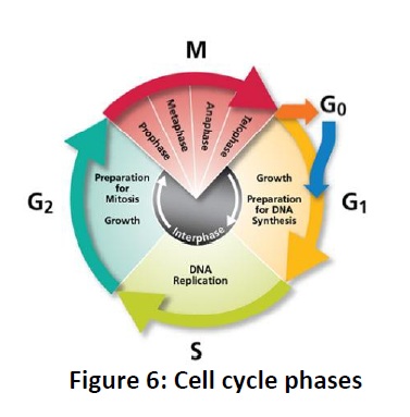
Cell Cycle:
The
cell cycle is made up of four phases (Fig. 6). In the M phase (M=mitosis), the
chromatin condenses into chromosomes, and the two individual chromatids, which
make up the chromosome, segregate to each daughter cell. In the G1 (Gap 1)
phase, the cell either progresses toward DNA synthesis or another division
cycle or exits the cell cycle reversibly (G0) or irreversibly to commit to
differentiation. During G1, the cell is particularly susceptible to control of
cell cycle progression; this may occur at a number of restriction points, which
determine whether the cell will re-enter the cycle, withdraw from it, or
withdraw and differentiate. G1 is followed by the S phase (DNA synthesis), in
which the DNA replicates. S in turn is followed by the G2 (Gap 2) phase in
which the cell prepares for reentry into mitosis. Checkpoints, at the beginning
of DNA synthesis and in G2, determine the integrity of the DNA and will halt
the cell cycle to allow either DNA repair or entry into apoptosis if repair is
impossible. The Phospho Histone H3 Imaging Kit (Roche) is a convenient method
for fast cell cycle analysis by quantification of mitotic cells. Apoptosis, or
programmed cell death, is a regulated physiological process whereby a cell can
be removed from a population. Characterized by DNA fragmentation, nuclear
blebbing, and cell shrinkage, apoptosis can be detected via a number of marker
enzymes and kits (see Roche Applied Science products). Roche DNA Fragmentation
Imaging Kit is a TUNEL assay-based method for accurate and fast quantitative
fluorescence detection of apoptosis in medium to high throughput cellular workflows.
Related Topics