Chapter: Basic Concept of Biotechnology : Animal Biotechnology
Biological Contamination
Biological
Contamination
Contamination
of cell cultures is the common problem encountered in cell culture
laboratories, sometimes with very serious consequences. Cell culture
contaminants can be divided into two main categories, chemical contaminants such as impurities in media, sera, and water,
endotoxins, plasticizers, and detergents, and biologicalcontaminants such as bacteria, molds, yeasts, viruses,
mycoplasma, aswell as cross contamination by other cell lines. While it is
impossible to eliminate contamination entirely, it is possible to reduce its
frequency and seriousness by gaining a thorough understanding of their sources
and by following good aseptic technique. This section provides an overview of
major types of biological contamination. Bacterialcontamination is easily
detected by visual inspection of the culture within a few days of it becoming
infected; infected cultures usually appear cloudy (i.e., turbid), sometimes
with a thin film on the surface. Sudden drops in the pH of the culture medium
are also frequently encountered. Under a low power microscope, the bacteria
appear as tiny, moving granules between the cells, and observation under a
high-power microscope can resolve the shapes of individual bacteria. The
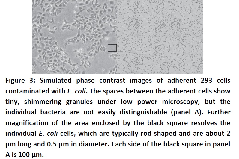
simulated images below show an adherent 293 cell culture contaminated with E. coli

Figure 3: Simulated phase contrast
images of adherent 293 cells contaminated with E. coli. The spaces between the adherent cells show tiny,
shimmering granules under low power microscopy, but the individual bacteria are
not easily distinguishable (panel A). Further magnification of the area
enclosed by the black square resolves the individual E. coli cells, which are typically rod-shaped and are about 2 μm
long and 0.5 μm in diameter. Each side of the black square in panel A is 100
μm.
Yeasts
are unicellular eukaryotic microorganisms in the kingdom of Fungi, ranging in
size from a few micrometers (typically) up to 40 micrometers (rarely). Like
bacterial contamination, cultures contaminated with yeasts become turbid,
especially if the contamination is in an advanced stage. There is very little
change in the pH of the culture contaminated by yeasts until the contamination
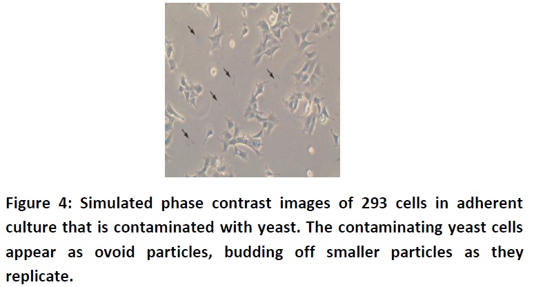
becomes heavy,at which stage the pH usually increases. Under microscopy, yeast
appears as individual ovoid or spherical particles, which may bud off smaller
particles. The simulated image below shows adherent 293 cell culture 24 hours
after plating that is infected with yeast (Fig. 4).

Similar to yeast contamination, the pH of the
culture remains stable in the initial stages of contamination, then rapidly
increases as the culture become more heavily infected and becomes turbid. Under
microscopy, the mycelia usually appear as thin, wisp-like filaments, and
sometimes as denser clumps of spores. Spores of many mold species can survive extremely
harsh and inhospitable environments in their dormant stage, only to become
activated when they encounter suitable growth conditions. Viruses are
microscopic infectious agents that take over the host cells machinery to
reproduce. Their extremely small size makes them very difficult to detect in
culture, and to remove them from reagents used in cell culture laboratories.
Because most viruses have very stringent requirements for their host, they
usually do not adversely affect cell cultures from species other than their
host. However, using virally infected cell cultures can present a serious
health hazard to thelaboratory personnel, especially if human or primate cells
are cultured in the laboratory. Viral infection of cell cultures can be
detected by electron microscopy, immunostaining with a panel of antibodies,
ELISA assays, or PCR with appropriate viral primers.
Related Topics