Chapter: Biotechnology Applying the Genetic Revolution: Biowarfare and Bioterrorism
Biosensors and Detection of Biowarfare Agents
BIOSENSORS
AND DETECTION OF BIOWARFARE AGENTS
Biosensors are devices for detection and
measurement of reactions that rely on a biological mechanism, often an enzyme
reaction adapted to generate an electrical signal. Biosensors have been traditionally
used in clinical diagnosis and in food and environmental analysis. By far the
biggest use has been the clinical monitoring of glucose levels using the enzyme
glucose oxidase. In particular, knowing the concentration of glucose is critical
to proper care of diabetics. Glucose oxidase is unusually stable, a major reason
for its widespread use. The enzyme catalyzes the following reaction:
glucose +O 2 → - gluconolactone +H 2 O 2

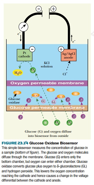
The glucose biosensor consists of a thin
layer of glucose oxidase attached to the bottom of an oxygen electrode ( Fig.
23.14 ). The electrode detects oxygen released by the enzyme reaction. The
current generated provides a measure of the glucose concentration. A potential
of about 0.6 volts is applied between the central positive platinum electrode
and the surrounding negative silver/silver chloride electrode. The electrolyte
solution is saturated potassium chloride. The negative electrode (cathode) is
covered by a thin Teflon membrane, which allows oxygen to diffuse through but
keeps out other molecules that might react. The electrode reactions are:
Platinum cathode ( electrons consumed )
O2 4H ++ 4 e − → 2
H 2 O
Ag / AgCl anode ( electrons released )
4Ag 4Cl − → 4 AgCl +4 e −
There is growing interest today in using
biosensors to detect toxins, viruses, and perhaps other possible biowarfare
agents. In particular a handheld device giving a rapid response would be highly
useful. Several proposals exist that would use specific antibodies or antibody
fragments as detectors for biowarfare agents. Each B cell carries antibodies
specific for one antigen and one proposal is to use whole B cells in the
biosensor. When an antigen binds to the antibody on the surface of a B cell,
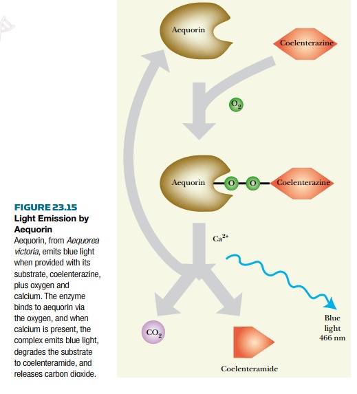
it triggers a signal cascade. Engineered
B cells have been made that express aequorin , a light-emitting protein from
the luminescent jellyfish Aequorea victoria . Aequorin emits blue light
when triggered by calcium ions ( Fig. 23.15 ). Living jellyfish actually
produce flashes of blue light, which are transduced to green by the famous
green fluorescent protein (GFP).

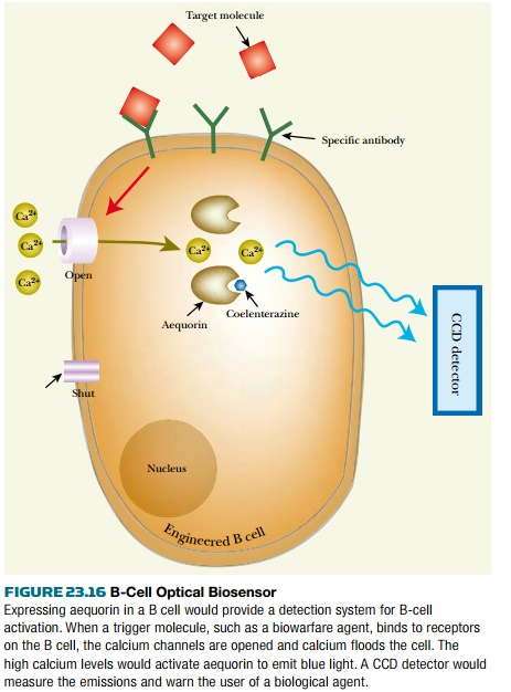
In the biosensor, when a B cell detected
a disease agent (or any targeted microbe), the cell would release calcium ions
due to activation of a signal cascade ( Fig. 23.16 ). This in turn triggers
light emission by aequorin. The light emitted is detected by a sensitive
chargecoupled device (CCD) detector. This approach is in its developmental
stages and will allow detection of 5 to 10 particles of a pathogenic agent such
as a virus or bacterium. Multiple patches of around 10,000 B cells specific to
different pathogens would be assembled in array fashion onto the same chip.
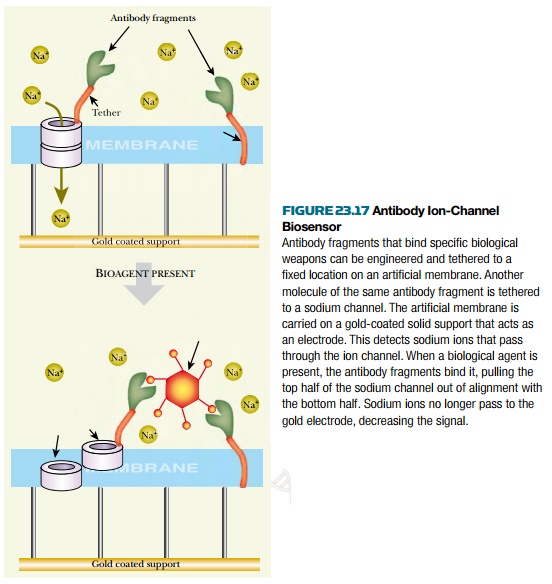
Another scheme, under development by the
Ambri Corporation of Australia, uses antibody fragments mounted on an
artificial biological membrane, which is attached to a solid support covered by
a gold electrode layer. Channels for sodium ions are incorporated into the
membrane. When the ion channels are open, sodium ions flow across the
membraneand a current is generated in the gold electrode. The ion channels
consist of two modules,each spanning half the membrane. When top and bottom
modules are united, the ion channel is open. When the top module is pulled
away, the ion channel cannot operate. Binding of biowarfare agents by the
antibody fragments separates the two halves of the channels, which in turn
affects the electrical signal ( Fig. 23.17 ).


Related Topics