Chapter: Medical Surgical Nursing: Assessment of Respiratory Function
Anatomy of the Upper Respiratory Tract
Anatomic and Physiologic Overview
The respiratory system is composed of
the upper and lower res-piratory tracts. Together, the two tracts are
responsible for ven-tilation (movement
of air in and out of the airways). The uppertract, known as the upper airway,
warms and filters inspired air so that the lower respiratory tract (the lungs)
can accomplish gas exchange. Gas exchange involves delivering oxygen to the
tissues through the bloodstream and expelling waste gases, such as car-bon
dioxide, during expiration.
ANATOMY OF THE UPPER RESPIRATORY TRACT
Upper airway structures consist of the
nose, sinuses and nasal pas-sages, pharynx, tonsils and adenoids, larynx, and
trachea.
Nose
The
nose is composed of an external and an internal portion. The external portion
protrudes from the face and is supported by the nasal bones and cartilage. The
anterior nares (nostrils) are the ex-ternal openings of the nasal cavities.
The internal portion of the nose is a
hollow cavity separated into the right and left nasal cavities by a narrow
vertical divider, the septum. Each nasal cavity is divided into three
passageways by the projection of the turbinates (also called conchae) from the lateral
walls. The nasal cavities are lined with highly vascular cil-iated mucous
membranes called the nasal mucosa. Mucus, se-creted continuously by goblet
cells, covers the surface of the nasal mucosa and is moved back to the
nasopharynx by the action of the cilia
(fine hairs).
The
nose serves as a passageway for air to pass to and from the lungs. It filters
impurities and humidifies and warms the air as it is inhaled. It is responsible
for olfaction (smell) because the ol-factory receptors are located in the nasal
mucosa. This function diminishes with age.
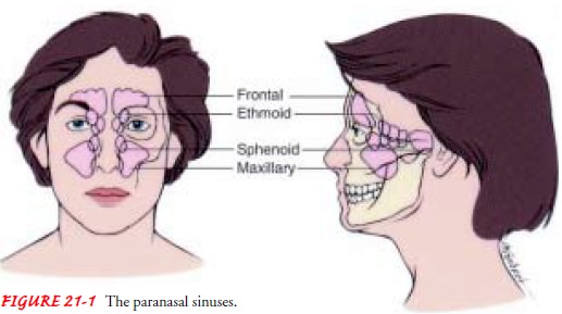
Paranasal Sinuses
The
paranasal sinuses include four pairs of bony cavities that are lined with nasal
mucosa and ciliated pseudostratified columnar epithelium. These air spaces are
connected by a series of ducts that drain into the nasal cavity. The sinuses
are named by their location: frontal, ethmoidal, sphenoidal, and maxillary
(Fig. 21-1). A prominent function of the sinuses is to serve as a resonating
chamber in speech. The sinuses are a common site of infection.
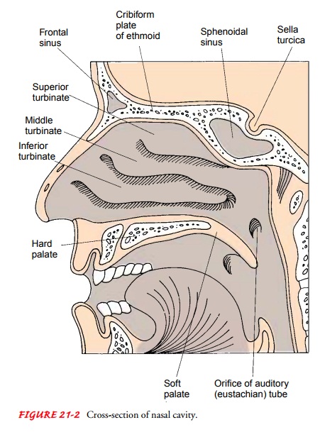
Turbinate Bones (Conchae)
The
turbinate bones are also called conchae (the name suggested by their shell-like
appearance). Because of their curves, these bones increase the mucous membrane
surface of the nasal passages and slightly obstruct the air flowing through
them (Fig. 21-2).
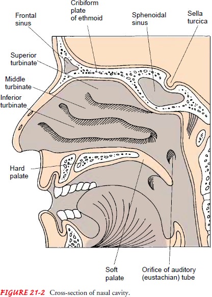
Air
entering the nostrils is deflected upward to the roof of the nose, and it
follows a circuitous route before it reaches the na-sopharynx. It comes into
contact with a large surface of moist, warm mucous membrane that catches
practically all the dust and organisms in the inhaled air. The air is
moistened, warmed to body temperature, and brought into contact with sensitive
nerves. Some of these nerves detect odors; others provoke sneezing to expel
irritating dust.
Pharynx, Tonsils, and Adenoids
The
pharynx, or throat, is a tubelike structure that connects the nasal and oral
cavities to the larynx. It is divided into three re-gions: nasal, oral, and
laryngeal. The nasopharynx is located pos-terior to the nose and above the soft
palate. The oropharynx houses the faucial, or palatine, tonsils. The
laryngopharynx ex-tends from the hyoid bone to the cricoid cartilage. The
epiglottis forms the entrance of the larynx.
The
adenoids, or pharyngeal tonsils, are located in the roof of the nasopharynx.
The tonsils, the adenoids, and other lymphoid tissue encircle the throat. These
structures are important links in the chain of lymph nodes guarding the body
from invasion by or-ganisms entering the nose and the throat. The pharynx functions
as a passageway for the respiratory and digestive tracts.
Larynx
The larynx, or voice organ, is a cartilaginous epithelium-lined structure that connects the pharynx and the trachea. The major function of the larynx is vocalization. It also protects the lower airway from foreign substances and facilitates coughing. It is fre-quently referred to as the voice box and consists of the following:
·
Epiglottis—a valve flap of cartilage
that covers the opening to the larynx during swallowing
·
Glottis—the opening between the
vocal cords in the larynx
·
Thyroid cartilage—the largest of the
cartilage structures; part of it forms the Adam’s apple
·
Cricoid cartilage—the only complete
cartilaginous ring in the larynx (located below the thyroid cartilage)
·
Arytenoid cartilages—used in vocal
cord movement with the thyroid cartilage
·
Vocal cords—ligaments controlled by
muscular movements that produce sounds; located in the lumen of the larynx
Trachea
The
trachea, or windpipe, is composed of smooth muscle with C-shaped rings of
cartilage at regular intervals. The cartilaginous rings are incomplete on the
posterior surface and give firmness to the wall of the trachea, preventing it
from collapsing. The trachea serves as the passage between the larynx and the
bronchi.
Related Topics