Chapter: Human Nervous System and Sensory Organs : The Ear
Vestibular Apperatus - Structure of The Ear
Vestibular Apperatus
Saccule, utricle, and the three semicircularducts emanating from the
utricle form theorgan of balance, the vestibular
apparatus. It contains several sensory fields, namely, the two acoustic
maculae, macula of saccule and macula of utricle, and the three ampullarycrests. They all register
acceleration andpositional changes and, therefore, serve spatial orientation.
The maculae react to linear acceleration in different directions, the crests
react to rotational acceleration. The maculae occupy specific positions in
space; the macula of the utricle lies roughly horizontally on the floor of the
utricle, and the macula of the saccule lies vertically on the anterior wall of
the sac-cule. Thus, both are arranged at right angles to each other. (For
positions of the semi-circular ducts, )
Acoustic maculae (A).The epithelium lining the endolymphatic space increases
in height in the oval areas of the maculae and differentiates into
supporting cells and sensory cells. The supporting
cells (A1) carry and surround
the sensory cells (A2). Each sensory cell is shaped like a
flask or am-poule and bears 70 – 80 stereovilli on its api-cal surface (A3). The sensory epithelium is
surmounted by a gelatinous membrane, the statoconic
(otolithic) membrane (A4),
whichcarries crystalline particles of calcium car-bonate, the ear crystals, or statocones(otoliths) (A5). The stereovilli of the
sensorycells do not directly project into the stato-conic membrane but are
surrounded by a narrow space containing endolymph.
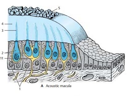
Function of the maculae. The
properstimulus for the stereovilli is a shearing force affecting the macula;
with increasing acceleration there is
a tangential shift be-tween sensory epithelium and statolithic membrane. The
resulting deflection of the stereovilli leads to stimulation of the sensory
cell and to induction of a nerve im-pulse.
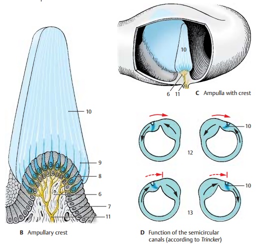
Ampullary crest (B, C). The crest (BC6) isformed by a protrusion in the ampulla and is oriented
transversely to the course of the semicircular duct (C). Its surface is covered by supporting
cells (B7) and sensory cells(B8). Each sensory cell bears approximately 50 stereovilli (B9) that are considerably longer than
those of the macular cells. The ampullary crest occupies about one third of the
height of the ampulla. It is surmounted by a gelatinous cap, the ampullarycupula (B – D10), which reaches
to the roof of the ampulla. The cupula is traversed by long channels into which
the hair bundles of the sensory cells protrude. The bases of the sensory cells
are innervated by nerve endings (A –
C11).

Function of the semicircular canals (D).
The
semicircular canals respond to ro-tational
acceleration which sets the en-dolymph in motion. The resulting deflection
of the ampullarycupula bends the stereovilli of the sensory cells and acts as
the triggering stimulus. For example, if the head is turned to the right (red
arrows), the endolymph of the lateral semicircular duct initially remains in
place due to its inertia; this results in a relative movement in the opposite
direction (hydrodynamic inertia, black arrows) so that both cupulae are de-flected
toward the left (D12). The
en-dolymph then slowly follows the rotation of the head. However, once the
rotation has stopped (broken, arrested arrows), it con-tinues to flow for a
certain distance in the same direction so that the cupulae are de-flected to
the right (D13). The function of the
semicircular ducts serves primarily the reflex eye movements. Rapid eye
move-ments caused by rotation of the head (ro-tatorynystagmus)
depend on cupular de-flection. The slow component of nystagmus always follows
the direction of the cupular deflection.
Related Topics