Chapter: Human Nervous System and Sensory Organs : Basic Elements of the Nervous System
Ultrastructure of the Nerve Cell
Ultrastructure of the Nerve Cell
Electron micrographs show the cell nucleus (A – C1) to be enclosed
by a double-layeredmembrane (A2). It contains the nuclear pores (BC3) that probably open only temporarily. The karyoplasm of the nucleus contains finely dispersed chromatin granules, which consist of DNA
and proteins. The nucleolus (A – C4), a spongiform area of the nucleus made up of a dense granular
component and a loose filamentous component, con-sists of RNA and proteins.
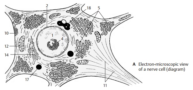
In the cytoplasm, the Nissl bodies appear as rough endoplasmic reticulum (A
– C5), a lamel-lar system of
membranes that enclose flat-tened, intercommunicating cisternae (BC6). Attached to the cytoplasmic side
of the membranes are the protein-synthesizing ri-bosomes (BC7). To
maintain the long axon(up to 1 m long), it is essential that the cell has an
extremely high rate of protein syn-thesis (structural metabolism).
Ribosome-free membranes form the agranular or smooth endoplasmic reticulum (C8).
The roughendoplasmic reticulum communicates with the perinuclear space (BC9)
and with the marginal cisternae (A10) below the cell sur-face. Marginal
cisternae are often found at sites where boutons or glial cell processes are
attached. The cytoplasm is crossed by neurofilaments
and neurotubules (A –
C11)that are arranged into long parallel bundles inside the axon. The
neurotubules correspond to the microtubules of other cells.
The transport of substances takes
place along neurofilaments and neurotubules ( D). Neurofibrils are the light-micro-scopic equivalent of densely
packed neu-rotubules.
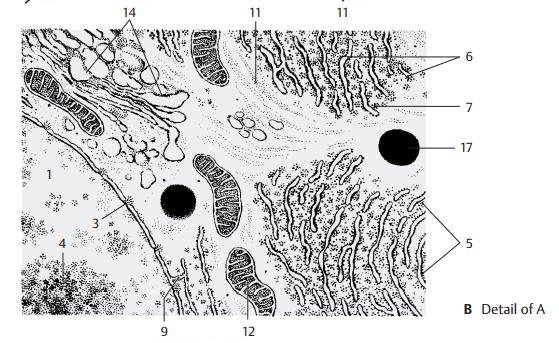
The neuron contains a large number
of mito-chondria (A –
C12). These are enclosed in adouble membrane; the inner membrane shows
projections (cristae) (C13) into the inner space (matrix). The mitochondria are of various
shapes (short and plump in the perikaryon, long and slender in the den-drites
and the axon) and move constantly along fixed cytoplasmic paths between
theNissl bodies. The mitochondria are the site of cellular respiration and,
hence, of energy generation. Numerous enzymes are local-ized in the inner
membrane and in the matrix, among others the enzymes of the citric acid cycle and respiratory-chain (oxida-tive)
phosphorylation.
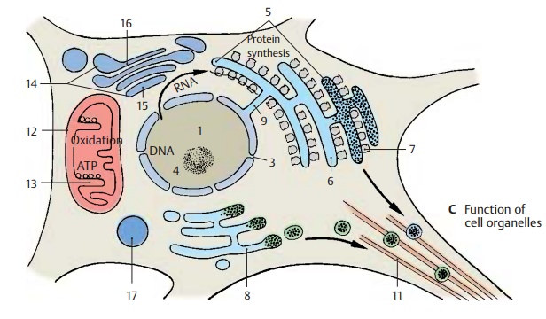
The Golgi complex consists of a number of dictyosomes (A – C14), which are stacks offlattened,
noncommunicating cisternae. The dictyosome has a forming side (cis face) (C15) and a maturing side (trans
face) (C16). The forming side
receives transport vesicles from the endoplasmic reticulum. At the margins of
the maturing side, Golgi vesicles are
formed by budding. The Golgi complex is mainly involved in the modification
(e.g., glycosylation, phosphorylation) of proteins from the endoplasmic
reticulum.
The numerous lysosomes (A – C17) contain various enzymes (e.g.,
esterases, proteases) and are mainly involved in intracellular digestion.
A18 Pigment.
Related Topics