Chapter: Human Nervous System and Sensory Organs : The Eye
Topographic Organization of the Visual Pathway - Eye
Topographic Organization of the Visual Pathway
The
fibers from individual regions of the retina occupy specific positions in the
different parts of the optic system. To illustrate this in a simple way, the
retina is subdivided into four quadrants that all share the center, namely, the
macula with the central fovea (area of highest visual acuity). The fibers of
the fovea show a regular point-to-point connection between fovea, lateral
genicu-late body, and striate area.
The
halves of the visual fields of each eye (visual
hemifields) (A1) are projected
onto the respective contralateral halves of the re-spectiveretina (hemiretinas) (A2). Immedi-ately after the exit of the
optic nerve from the eyeball (A3), the macular fibers lie on the
lateral side of the nerve, with the fibers from the nasal half of the macula
lying in the cen-ter, surrounded by fibers of the temporal half of the macula.
Further along the optic nerve, the macular bundle comes to occupy the center (A4).
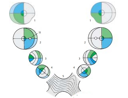
The
fibers of the nasal hemiretinas (con-tinuous lines) cross to the opposite side
in the optic chiasm (A5). While doing so they take a strange
course. The medial fibers cross, then
run a short distance into the con-tralateral optic nerve, and finally turn in a
right angle into the contralateral optic tract. The lateral fibers run a short distance into the ipsilateral optic
tract and then turn abruptly into the contralateral tract. The fibers of the
temporal hemiretinas (broken lines) do not cross but continue in the
ipsi-lateral tract.
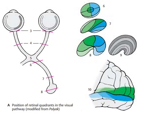
The optic tract (A6) thus contains the fibers of the corresponding halves of both
retinas: the left tract contains the fibers of the left hemiretinas, the right
tract contains the fibers of the right hemiretinas. The fibers of the two
superior retinal quadrants lieven-tromedially, those of the two inferior
quad-rants lie dorsolaterally, while the fibers of the macula take a central
position. Prior to radiating into the lateral geniculate body (A7), the fibers rearrange so that the
macu-lar fibers form a central wedge, the fibersfrom the upper retinal
quadrants come to lie medially, and the fibers from the lower reti-nal
quadrants lie laterally.
The
fibers in the lateral geniculate body
(A8) terminate in the same
arrangement. The central wedge of the terminal macular fibers makes up almost
half of the genicu-late body. The fibers from the periphery of the retina
terminate in the most anterior and ventral regions of the lateral geniculate
body. The terminals of the ipsilateral and contralateral fibers in the
geniculate layers are shown schematically in light gray and dark gray (A9) (see also p. 257, A). The
geniculate cells of the central wedge project to the posterior region of the striate area (A10). The area of highest acuity, which in the human retina
measures slightly more than 2 mm in diameter, is represented by the largest
portion of the visual cortex. Ros-trally to it lie the much smaller areas for
the remaining parts of the retina. The upper quadrants of each retina are
represented in the upper lip of the calcarine sulcus, and the lower quadrants
in the lower lip.
Clinical Note: Corresponding to the arrange-ment
of fibers, injury to the visual pathway in specific segments results in various
patterns of loss of vision. It should be taken into considera-tion that the
lower halves of each retina register the input from the upper halves of the
visual fields, while the upper halves of each retina regis-ter the input from
the lower halves of the visual fields. The same is true for the left and right
halves of each retina. Injury to the optic tract, lateral geniculate body, or
visual cortex on the left side affects the left halves of each retina and the
right halves of each visual field. The result is ho-monymous hemianopia on the
right side. In case of bitemporal heteronymous hemianopia, injury to the
crossing nasal fibers of both retinae (e.g., in case of tumors of the
hypophysis near the optic chiasm) results in bilateral loss of the temporal
halves of the visual fields. Damage of both visual cortices causes visual
agnosia.
A11
Blind spot (papilla of the optic nerve).
Related Topics