Chapter: Basic Radiology : Imaging of the Heart and Great Vessels
Techniques and Normal Anatomy : Radionuclide Imaging (Nuclear Medicine)
Radionuclide
Imaging (Nuclear Medicine)
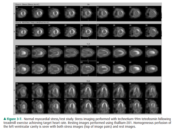
Cardiac radionuclide imaging,
primarily used for the patient with suspected myocardial ischemia or
infarction, requires an intravenous injection of radioactively labeled
compounds that have an affinity for the myocardium. These compounds localize
within the myocardium in diseased or damaged areas, and a radioactivity
detector such as a gamma camera can image their distribution. These tests are
most commonly used in the evaluation of patients with angina and atypical chest
pain (Figure 3-7). Gallium scans are occasionally used to assess for intrinsic
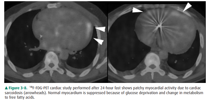
myocardial disease such as myocardial sarcoidosis. Positron emission tomography
(PET) with 18F-FDG (18F-fluorodeoxyglucose) is a
problem-solving tool that has shown promise in assessing myocardial viability
in pa-tients with known coronary artery disease and to assess for metabolically
active infiltrative disease (Figure 3-8). In addi-tion, rubidium-82 and
nitrogen-13 ammonia have been used as PET agents to evaluate myocardial
perfusion.
Related Topics