Chapter: Essentials of Anatomy and Physiology: Muscular System
Skeletal Muscle Structure
Skeletal Muscle Structure
Connective Tissue Coverings of Muscle
Each skeletal muscle is surrounded by a connective tissue sheath called the epimysium (ep-i-mis′ē-ŭm), or muscular fascia (fash′ē-ă) (figure 7.2a). Each whole muscle is subdivided by a loose con-nective tissue called the perimysium (per′ i-mis′ ē-ŭm) into numerous visible bundles called muscle fasciculi (fă-sik′ ū-lı̄). Each fascicle is then subdivided by a loose connective tissue called the endomysium (en′ dō-mis′ ē-ŭm) into separate muscle cells, called muscle fibers (figure 7.2b).
Muscle Fiber Structure
Examining the structure of a muscle fiber helps us understand the mechanism of muscle contraction. A muscle fiber is a single cylindrical fiber, with several nuclei located at its periphery. The largest human muscle fibers are up to 30 cm long and 0.15 mm in diameter. Such giant cells may contain several thousand nuclei. The cell membrane of the muscle fiber is called the sarcolemma(sar′kō-lem′ă; sarco, flesh) (figure 7.2b). The multiple nuclei of the muscle fiber are located just deep to the sarcolemma. Along the surface of the sarcolemma are many tubelike invaginations, called transverse tubules, or T tubules,which occur at regular intervals along the muscle fiber and extend inward into it.
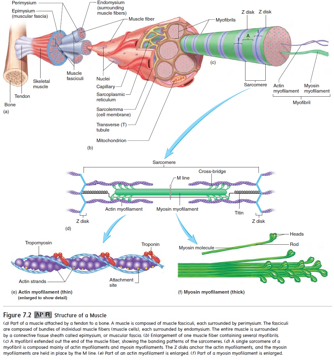
Figure 7.2 Structure of a Muscle
The T tubules are associated with a highly organized smooth endoplas-mic reticulum called the sarcoplasmic reticulum (re-tik′ū-lŭm). T tubules connect the sarcolemma to the sarcoplasmic reticulum. The sarcoplasmic reticulum has a relatively high concentration of Ca2+, which plays a major role in muscle contraction.
Inside each muscle fiber is the cytoplasm, called the sarcoplasm (sar′kō-plazm). It contains numerous myofibrils (mı̄-ō-f ı̄′brilz; myo, muscle), threadlike structures that extend from one end of the muscle fiber to the other (figure 7.2c). Myofibrils consist of two major kinds of protein fibers: actin (ak′ tin) myofilaments (mı̄-ō-fil′ ă-ments) and myosin (mı̄′ ō-sin) myofilaments (figure 7.2d). The actin and myosin myofilamentsare arranged into highly ordered, repeating units called sarco-meres (sar′kō-mērz), which are joined end-to-end to form themyofibrils (figure 7.2c,d).
Actin and Myosin Myofilaments
Actin myofilaments, or thin filaments, are made up of three com-ponents: actin, troponin, and tropomyosin. The actin strands, which resemble two minute strands of pearls twisted together, have attach-ment sites for the myosin myofilaments (figure 7.2e). Troponin (trō′pō-nin) molecules are attached at specific intervals along the actin myofilaments. These molecules have binding sites for Ca2+. Tropomyosin(trō-pō-mı̄′ō-sin) filaments are located along thegroove between the twisted strands of actin myofilament subunits. The tropomyosin filaments block the myosin myofilament binding sites on the actin myofilaments in an unstimulated muscle. In other words, if no Ca2+ is present, the tropomyosin filaments cover the attachment sites on the actin myofilament. However, when Ca2+ is present, it binds to troponin, which causes the tropomyosin fila-ments to expose the attachment sites on the actin myofilaments.
Myosin myofilaments, or thick myofilaments, resemble bundles of minute golf clubs (figure 7.2f ). The parts of the myosin molecule that resemble golf club heads are referred to as myosinheads. The myosin heads have three important properties: (1) Theheads can bind to attachment sites on the actin myofilaments;(2) they can bend and straighten during contraction; and (3) they can break down ATP, releasing energy.
Sarcomeres
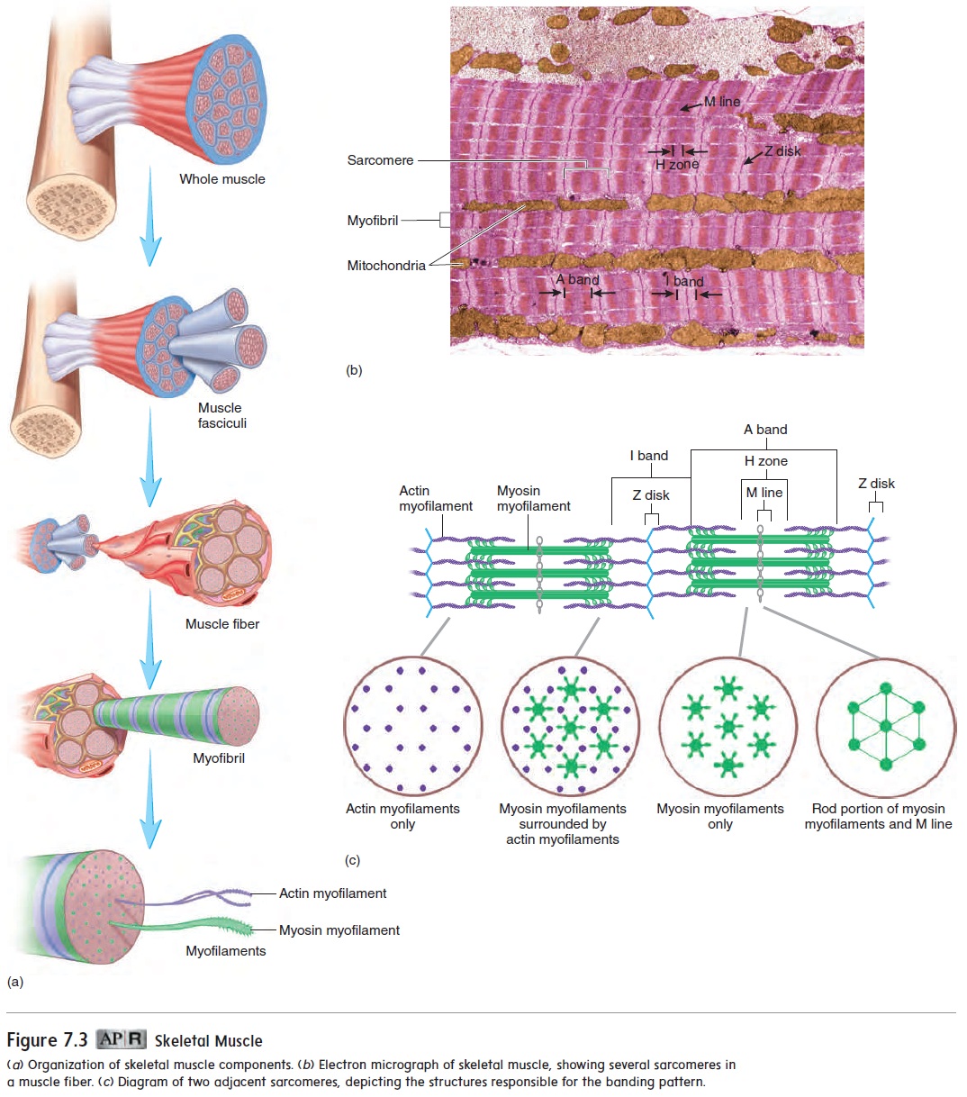
The sarcomere is the basic structural and functional unit of skel-etal muscle because it is the smallest portion of skeletal muscle capable of contracting. The separate components of the sarcomere can slide past each other, causing the sarcomeres to shorten. When the sarcomeres shorten, the myofibrils shorten, which is the ulti-mate cause of contraction of the muscle fiber during a contraction. Each sarcomere extends from one Z disk to an adjacent Z disk. Each Z disk is a network of protein fibers forming an attachment site for actin myofilaments. The arrangement of the actin and myosin myofilaments in sarcomeres gives the myofibril a banded appearance (figure 7.3). A light I band, which consists only of actin myofilaments, spans each Z disk and ends at the myosin myofilaments. A darker, central region in each sarcomere, called an A band, extends the length of the myosin myofilaments. The actin and myosin myofilaments overlap for some distance at both ends of the A band. In the center of each sarcomere is a second light zone, called the H zone, which consists only of myosin myofilaments. The myosin myofilaments are anchored in the center of the sarcomere at a dark-staining band, called the M line. The alternating I bands and A bands of the sarcomeres are respon-sible for the striations in skeletal muscle fibers observed through the microscope (see table 4.10a). It is the close association of the sarcomeres, the T tubules, and the sarcoplasmic reticulum that enables a nerve stimulus to initiate contraction of the muscle fiber.
Related Topics