Chapter: Human Nervous System and Sensory Organs : Brain Stem and Cranial Nerves
Pons
Pons
The semi-schematic cross sections show the cellular stain (Nissl) on the left and the corresponding fiber stain (myelin) on the right.
Cross Section at the Level of the Genu of the Facial Nerve (A)
Beneath the floor of the rhomboid fossa lies the magnocellular nucleus of the abducensnerve (A1) and, ventrolaterally to it, the nucleus of the facial nerve (A2). Thevisceroefferent superior salivatory nucleus (A3) is seen between the abducens and fa-cial nuclei. The lateral field is occupied by the sensory terminal nuclei of the vestibularnerve and the trigeminal nerve, namely, the medial vestibular nucleus (Schwalbe’s nu-cleus) (A4), the lateral vestibular nucleus (Deiters’ nucleus) (A5), and the spinal nu-cleus of the trigeminal nerve (A6).
The fibers of the facial nerve bend around the abducens nucleus (A1) and form the fa-cial colliculus (A7). We distinguish an as-cending limb (A8) and, cranial to the il-lustrated section, a descending limb. The apex is the internal genu of the facial nerve (A9). The fibers of the abducens nerve (A10) descend through the medial field of the teg-mentum. The medial longitudinal fasciculus (AB11) is seen medially and the posteriorlongitudinal nucleus (Schütz’s bundle)(AB12) dorsally to the abducens nucleus. Deep in the pontine tegmentum run the central tegmental tract (AB13) and the spinothalamic tract (A14). Secondary fibersof the auditory pathway collect from the anterior cochlear nucleus as a wide fiber bundle, the trapezoid body (AB15); they cross ventral to the medial lemniscus (A16) to the opposite side where they ascend in the lateral lemniscus (B17). They synapse in part in the adjacent nuclei of the trapezoid body, namely, the anterior nucleus of thetrapezoid body (A18) and the posterior nu-cleus of the trapezoid body (superior olive)(AB19). The spinal tract of the trigeminalnerve (A20) lies in the lateral field.
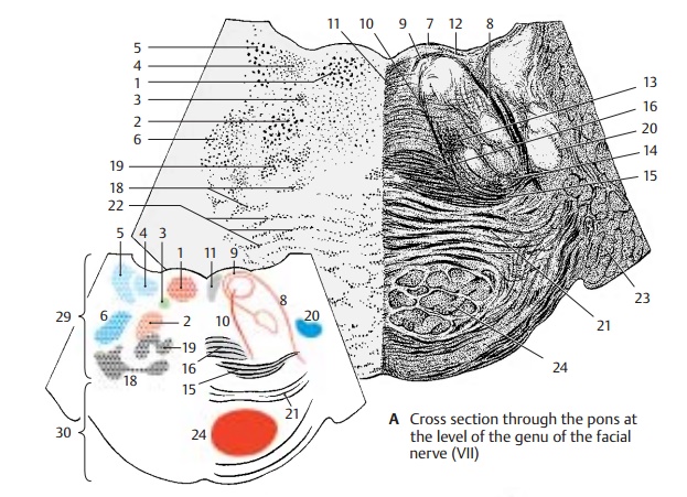
The pontine bulb is formed by the transversepontine fibers (A21). They are corticopon-tine fibers, which synapse in the pontine nu-clei (A22), and pontocerebellar fibers, whichare postsynaptic and extend to the cerebel-lum in the medial cerebellar peduncle (brachium pontis) (A23). Embedded in the middle of the longitudinally cut fiber bundles is the transversely cut fiber bundle of the pyramidal tract (AB24).
Cross Section at the Level of the Trigeminal Nerve (B)
The medial field of the pontine tegmentum is occupied by the nuclei of the tegmentum. These nuclei, of which only the inferior cen-tral tegmental nucleus (B25) is well defined,belong to the reticular formation. In the lateral field, the trigeminal complex has reached its widest expansion; lateral is the pontine nucleus of the trigeminal nerve (principal nucleus) (B26), medial to it the motor nucleus of the trigeminal nerve (B27),and dorsal the nucleus of the mesencephalic trigeminal root (B28). Afferent and efferent fibers unite to form the strong trunk emerg-ing at the anterior aspect of the pons.
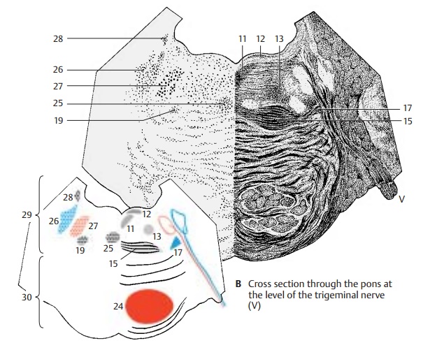
The lateral lemniscus (AB17), the trapezoidbody (AB15), and the adjacent posterior nu-cleus of the trapezoid body (AB19) lie ven-trally to the trigeminal nuclei. The following ascending and descending pathways can be recognized: the posterior longitudinalfasciculus (AB12), the medial longitudinal fasciculus (AB11), and the central tegmental tract (AB13).
AB29 Pontine tegmentum.
AB30 Pontine bulb.
Related Topics