Chapter: Essentials of Anatomy and Physiology: Muscular System
Nerve Supply and Muscle fiber Stimulation
Nerve Supply and Muscle fiber Stimulation
Skeletal muscle fibers do not contract unless they are stimulated by motor neurons. Motor neurons are specialized nerve cells that stimulate muscles to contract. Motor neurons generate action potentials that travel to skeletal muscle fibers. Axons of these neu-rons enter muscles and send out branches to several muscle fibers. Each branch forms a junction with a muscle fiber, called a neuro-muscular junction (figure 7.5). A more general term, synapse (sin′aps), refers to the cell-to-cell junction between a nerve cell and either another nerve cell or an effector cell, such as in a muscle or a gland. Neuromuscular junctions are located near the center of a muscle fiber. A single motor neuron and all the skeletal muscle fibers it innervates constitute a motor unit. A motor unit in a small, precisely controlled muscle, such as in the hand, may have only one or a few muscle fibers per unit, whereas the motor units of
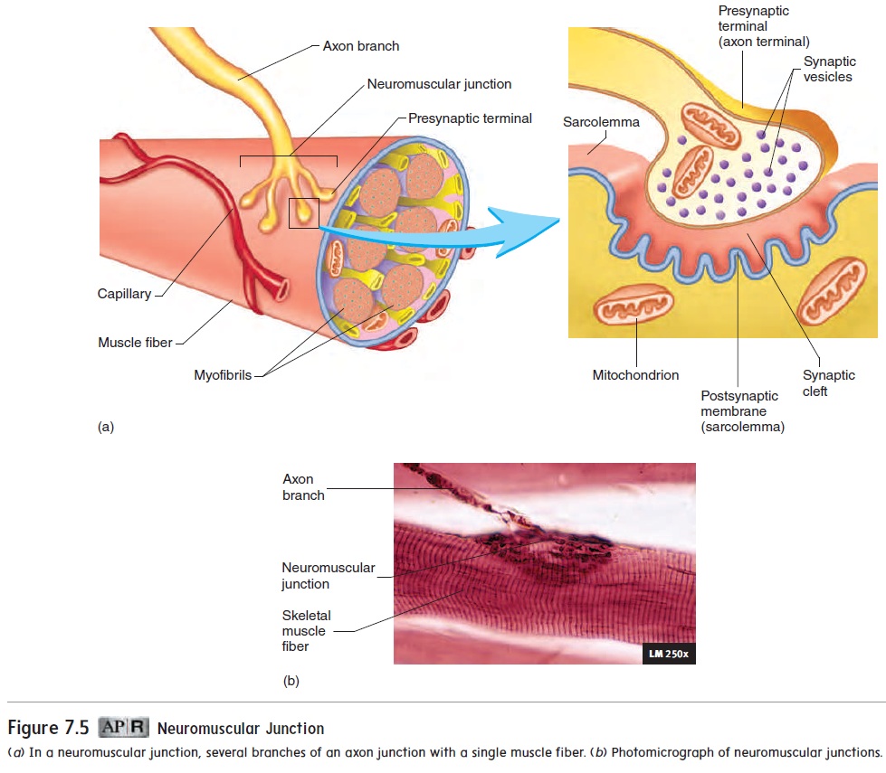
A neuromuscular junction is formed by a cluster of enlarged axon terminals resting in indentations of the muscle fiber’s cell membrane. An enlarged axon terminal is the presynaptic terminal; the space between the presynaptic terminal and the muscle fiber membrane is the synaptic cleft; and the muscle fiber membrane is the postsynaptic membrane. Each presynaptic terminal contains many small vesicles, called synaptic vesicles. These vesicles con-tain acetylcholine (as-e-til-kō′lēn), or ACh, which functions as aneurotransmitter, a molecule released by a presynaptic nerve cellthat stimulates or inhibits a postsynaptic cell.
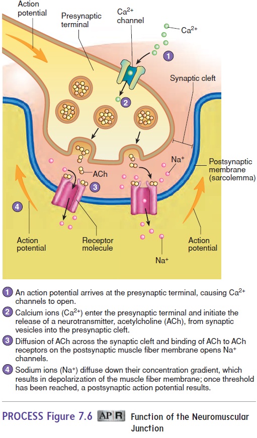
When an action potential reaches the presynaptic terminal, it causes Ca2+ channels to open. Calcium ions enter the presynaptic terminal and cause several synaptic vesicles to release acetyl-choline into the synaptic cleft by exocytosis (figure 7.6, steps 1 and 2). The acetylcholine diffuses across the synaptic cleft and binds to acetylcholine receptor sites on the Na+ channels in the muscle fiber cell membrane. The combination of acetylcholine with its receptor opens Na+ channels and therefore makes the cell membrane more permeable to Na+. The resulting movement of Na+ into the muscle fiber will initiate an action potential once threshold is reached. The action potential travels along the length of the muscle fiber and causes it to contract (figure 7.6, steps 3 and 4). The acetylcholine released into the synaptic cleft between the neuron and the muscle fiber is rapidly broken down by an enzyme, acetylcholinesterase(as′e-til-kō-lin-es′ter-ās). This enzymaticbreakdown ensures that one action potential in the neuron yields only one action potential in the skeletal muscle fibers of that motor unit and only one contraction of each muscle fiber.
Related Topics