Chapter: Human Nervous System and Sensory Organs : The Ear
Medial Wall of the Tympanic Cavity

Medial Wall of the Tympanic Cavity
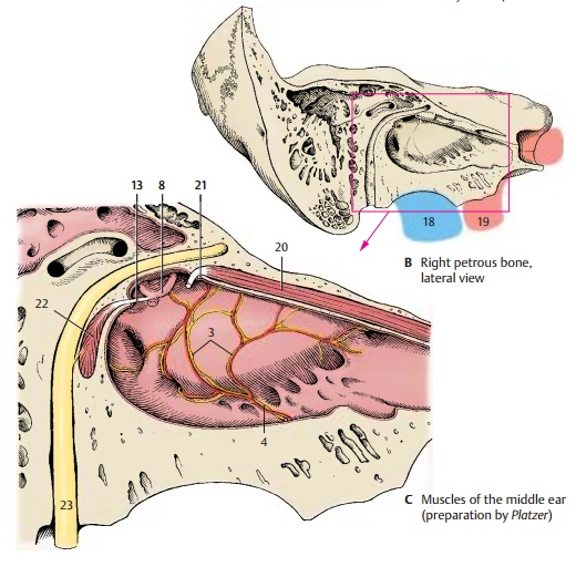
The medial wall, or labyrinthic wall, separates the tympanic cavity from the internal ear. The prominence in its middle region, the promontory of the tympanic cavity (A1), is caused by the basal convolution of the cochlea. In a bifurcated groove, the sulcus ofthe promontory (A2), lies the tympanic plexus (C3); it is formed by the tympanic nerve (C4) of the glossopharyngeal nerveand by the sympathetic fibers of the carotid plexus of the internal carotid artery. The promontory is anteriorly delimited by the tympanic cells (A5). In the medial wall, the oval window,vestibular window(A6), and the round window,cochlear window(A7), openinto the inner ear. The stapes (C8) rests in the vestibular window and closes it with its foot plate. The cochlear window is closed by the secondary tympanic membrane. In the posterior wall opening to the mastoid an-trum(A9) run two arched canals, the facial canal (A10) and the lateral semicircular canal (A11); both canals cause protrusionson the wall of the tympanic cavity, namely, the prominence of the facial canal and the prominence of the lateral semicircular canal.A bony protrusion, the pyramidal eminence (A12), contains an opening at its tip through which the tendon of the stapedius muscle (C13) enters. In anterior direction, the tym-panic cavity leads into the semicanal of theauditory tube (A14). Above it lies the semi-canal of the tensor tympani muscle (A15).The two semicanals are incompletely sepa-rated by a bony septum and together form the musculotubal canal. The medial wall at the level of the opening of the tympanic tube (carotid wall) separates the tympanic cavity from the carotid canal (A16); the bony floor (jugular wall) separates it from the jugular fossa (A17). Also shown are thejugu-lar vein (B18) and the internal carotid artery(B19).
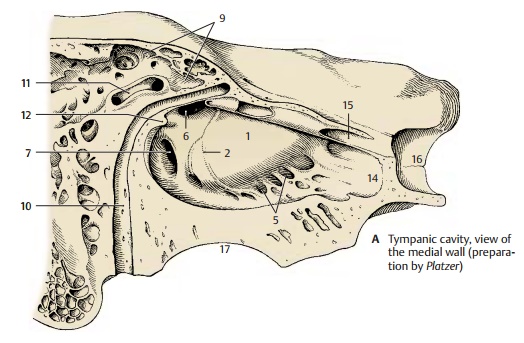
Clinical Note: The osseous roof and floor ofthe tympanic cavity may be very thin. In case of purulent otitis media, the infection can penetrate either through the roof and progress to meninges and brain (meningitis, cerebral abscess in thetemporal lobe), or through the floor and progressto the internal jugular vein (jugular thrombosis)
Related Topics