Chapter: BIOLOGY (ZOOLOGY) Standard XI first year 11th text book Assignment topics question and answer Explanation Definition
Male reproductive organs
Reproductive system
The process of sexual reproduction is a wonderful act in nature. This process, apart from ensuring a healthy progeny provides an oppurtunity to produce enormous range of genetically varied offsprings. Organisms have adopted several strategies for sexual reproductive processes. Such adaptations have resulted in suitable morphological, anatomical and behav-ioral modifications. Human reproductive organs as internal and external geni-talia are highly sophisticated yet simple in their functioning. The functioning is in accordance with psychological and endocrinological thresholds. An academic approach towards an understanding of the human male and female reproductive organs and their functions will go a long way in avoidance of unethical, unhealthy and unhygenic practices encoun-tered at specific periods in life.
Male reproductive organs
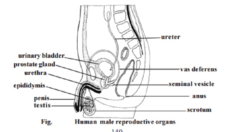
The male reproductive system consists of the testes (singular : tes-tis), epididymides (sing: epididymis), ductus deferentia or vasa deferentia
(sing : ductus deferens, vas deferens), urethra, seminal vesicles, prostate gland, bulbourethral glands, scrotum and penis.
Testes : The testes are the primary reproductive organs or gonads in the male. These are suspended in the scrotum by scrotal tissues.
The sperm cells are temperature sensitive. They do not develop nor-mally at usual body temperatures. Hence the testes and epididymides in which the sperm cells develop, are located outside body cavity in the scrotum, where the temperature is low.
The left testis usually is 1 cm lower than the right. An average testis is 4-5 cm in length, 2-5cm in breadth. Its weight varies from 10.5-14g.
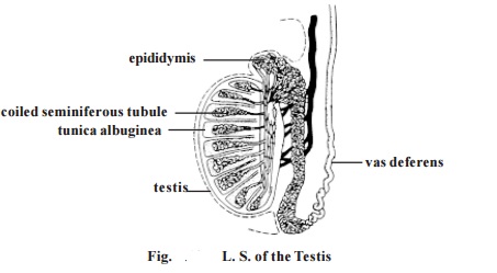
The outer part of each testis is a thick, white capsule called tunica albuginea. Internally the testis contains several incomplete septa. The septa divide each testis into about 300-400 cone shaped lobules. The lobules contain seminiferous tubules and interstitial cells or Leydig cells. Sperm cells de-velop within the seminiferous tubules.
The seminiferous tubules are extensive. The combined length of the tubules in both testes is nearly 800 metres. These tubules through a set of short, straight tubules open into tubular network called the rete testis. The rete testis in turn open into efferent ductules. Internally the tu-bules and ductules are lined by ciliated columnar epithelium. These cells help to move the sperm cells out of the testis.
Epididymis : It is formed of extremely convoluted ductules coming out of the testis. It occurs on the posterior side of the testis. The maturation of sperm cells occurs within the ductules of the epididymis.
Vas deferens or ductus deferens : It emerges from the tail end of the epididymis and ascends along the posterior side of the testis. It becomes asso-ciated with the blood vessels and nerves that supply the testis. Collectively these structures constitute the spermatic cord. Thus the spermatic cord con-sists of (1) Vas deferens (2) testicular artery and venus plexus (3) lymph vessels (4) nerves (5) fibrous processes and muscles. This cord enters into the pelvic region. The end of the vas deferns enlarges to form the ampulla. At this region the vas deferens is surrounded by smooth muscles capable of peri-staltic contraction. They help to propel the sperm cells through the ductus deferens.
Ejaculatory duct : Nearer to the ampulla of each vas deferens there is a sac like seminal vesicle. It joins the ductus deferens to form the ejaculatory duct. These ducts are about 2.5 cm long. They project into the prostate gland and end by opening into the urethra.
Urethra : The male urethra extends from the urinary bladder to the distal end of the penis. It is about 20 cm long. It is a passageway for both urine and reproductive fluids. The urethra is divided into three parts. They are
1. The prostatic Urethra - It is closest to the bladder and passes through the prostate gland.
2. The membranous urethra - It is the shortest part of the urethra and it extends from the prostatic urethra.
3. The spongy urethra or penile urethra - It is the longest part of the urethra. It extends from the membranous urethra, through the length of the penis. There are several minute mucus secreting urethral glands opening into the urethral passage.
Penis - It is the male copulatory organ. It consists of two parts namely the radix or root and the corpus or body. The radix attaches the penis to the lower abdomen. The corpus is normally pendulous. It is covered by a loose skin.
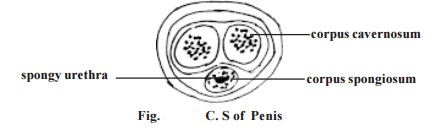
The corpus of the penis consists of three masses of erectile tissue. Flooding these tissues with blood causes the penis to enlarge and become firm. These tissues are the right and left corpora cavernosa and the median corpus spongiosum penis. Most of the corpus is formed of the corpora cavernoas. The corpus sporgiosum penis surrounds the urethra and near the end of the penis it expands into a conical, glans penis. Its swollen base is the corona glandis.
The skin over the penis is thin. It is loosely connected to the tunica albuginea. At the tip of the penis it is folded to form the prepuce or the foreskin. It overlaps the glans penis. The corona glandis and penile neck have numerous preputial glands.
Seminal vesicles - These are two sac-like structures located between the bladder and rectum. Each vesicle is about 5 cm long. Their secretions contrib-ute about 70% of the seminal fluid.
Prostate - It is a firm structure. It is partly glandular and partly fibromuscu-lar. It is found around the beginning of the male urethra. It is about 3 cm in diameter. It weighs about 8g.
The muscular part of the prostate may help in dilating the urethra to hold the seminal fluid (3-5ml) during the period of sexual excitement prior to ejaculation.
After the middle age the prostate often enlarges. It may project into the bladder and interrupt urination.
Bulbo-urethral gland - These are two glands. They are small round masses about 1 cm in diameter. They lie lateral to the membranous urethra. Its secre-tion may control genito-urinary diseases.
Scrotum - It is a fibromuscular sac. It contains the testes and their associated ducts. It is divided into right and left by cutaneous raphe. Its left side is usually lower. The external appearence varies according to age and body tempera-ture. The scrotal skin is thin and pigmented. It has numerous sweat glands and nerve endings.
Related Topics