Chapter: BIOLOGY (ZOOLOGY) Standard XI first year 11th text book Assignment topics question and answer Explanation Definition
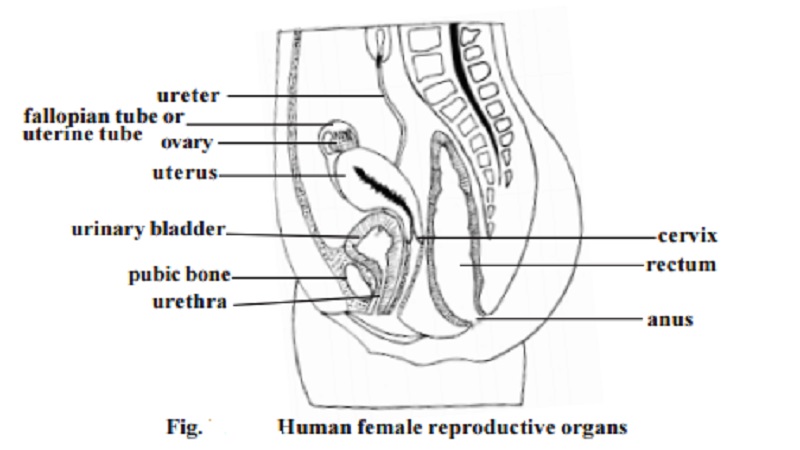
Female reproductive organs
Reproductive system
The process of sexual reproduction is a wonderful act in nature. This process, apart from ensuring a healthy progeny provides an oppurtunity to produce enormous range of genetically varied offsprings. Organisms have adopted several strategies for sexual reproductive processes. Such adaptations have resulted in suitable morphological, anatomical and behav-ioral modifications. Human reproductive organs as internal and external geni-talia are highly sophisticated yet simple in their functioning. The functioning is in accordance with psychological and endocrinological thresholds. An academic approach towards an understanding of the human male and female reproductive organs and their functions will go a long way in avoidance of unethical, unhealthy and unhygenic practices encoun-tered at specific periods in life.
In human female the internal reproductive organs are the ovaries, uterus, uterine tubes and vagina. Externally the organs are the mons pu-bis, labia majora and labia minora, clitoris and vestibular glands.
Ovaries - These are paired structures. The two ovaries are placed on each side of the uterus in the pelvic region. They are greyish pink in colour. Each ovary is almond shaped. They are about 3cm long,1.5cm wide and 1cm thick.
The ovary is attached to the posterior surface of the inner body wall by a membranous fold called the mesovarium. The ovary is further supported by suspensory and ovarian ligaments.
Ovarian structure - In young female the surface of the ovary is covered by a layer of ovarian surface epithelium. It consists of a single layer of cuboi-dal cells. Beneath the epithelium the ovary is surrounded by a tough coat named tunica albuginea. It is made of collagenous tissue.

The overy proper is divisible into two regions, namely the cortex and the medulla. The cortex region contains the ovarian follicles. The medulla is interior. It receives blood vessels and nerves at the hilum.
After puberty the cortex forms the major part of the ovary. It con-tains ovarian follicles and corpora lutea of various sizes. Their size de-pends on the stage of menstrual cycle or age. The cortex is filled with stroma composed of collagen. The follicles are embedded in the stroma.
Ovarian follicles
The formation of the female gamete has many different phases and it is complex. At birth, the primordial follicles are found in the superficial zone of the cortex . They contain primary oocytes (about 25mm in diameter). Each one of them is surrounded by a single layer of flat follicular cells. The follicles undergo changes as the female attains puberty. The vari-ous follicular stages are :
1. Primary follicle - The follicle cells are converted from squamous to cuboidal cells. The follicular membrane or membrana granulosa becomes multilay-ered. The oocyte increases in size. It has an outer thick layer called the zona pellucida. The follicular cells divide and form granulosa cells.
2.Secondary follicle - It is about 20�m thick. The granulosa cells surround the oocyte and form a mound of cells called the cumulus ovaricus. The inner and outer theca become prominent. The theca interna is well established.
3. Tertiary follicle - Only one follicle reaches the tertiary stage. It increases in size (2mm diameter). Now it is called the graffian follicle. The oocyte and ring of cells surrounding the oocyte (corona radiata) break away and float freely in the follicular fluid. Finally the wall of the follicle ruptures and the contents are released into the peritoneum.
The ovary of the foetus at 5 months gestation has 7 million oocytes. At birth the ovary of the child contains about 1 million oocytes. Due to further degeneration at the time of puberty only about 40,000 oocytes remain. Of the 40,000 oocytes only about 400 undergo ovulation during the reproductive years.
Corpus luteum - It is formed after ovulation. The walls of the empty follicle collapses and fold extensively. The granulosa cells of the theca externa get enlarged.
They are now termed as luteal cells. They secrete hormones. In pregnancy the corpus luteum persists. Otherwise, it degenerates after 10-12 days. The connective tissue cells get enlarged. It becomes white in colour and is now called as the corpus albicans. In course of time it shrinks and disappears.
Uterine tubes (Fallopian tubes) - There are two uterine tubes or oviducts, one on each side of the uterus. Each one is associated with a ovary. Each tube is about 10 cm length. The terminal part of the tube is enlarged to form the infundibulum. It opens into the peritoneal cavity. The opening is called the ostium. The uterine tube consists of three parts. The part nearer to the in-fundibulum is called the ampulla. It is the longest part. That part of the tube nearer to the uterus is called the isthmus. It is narrow. The tubular part enter-ing into the uterus is called the uterine or intramural part.
Uterus
It is a hollow thick walled muscular organ. It is pear shaped. It is about 7.5cm long and 5 cm wide. It weighs about 50g.
During pregnancy its weight may go upto 1kg. Its larger rounded part is called as the fundus. The narrower part is called as the cervix. The cervix is directed inferiorly. The middle part is the b o d y . The uterus continues as the cervical canal and opens into the vagina through a opening called the ostium.
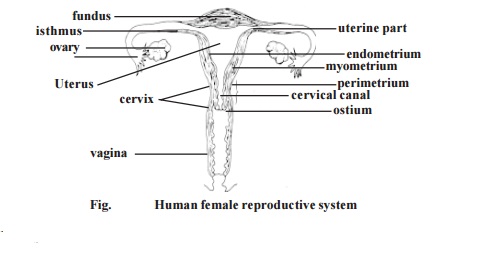
The wall of the uterus is three layered. The outermost layer is the perimetrium or serous layer. The major part of the wall is made up of the next layer called themyometrium or muscular coat . The innermost layer is the endometrium or mucous membrane. The endometrium is a functional layer. It undergoes menstrual changes and sloughing during female sex cycle.
Vagina - It is the female copulatory organ. It is a fibromuscular tube. It is about 10 cm long. It extends from the uterus to the outside. The vaginal pas-sage is used during intercourse and it allows menstrual flow and child birth.
External Genitalia
Vestibule - The external female genitalia is known as the vulva or puden-dum. It consists of the vestibule and its surrounding structures. The vestibular region remains in between the two labia majora. It contains the vaginal open-ing and the urethral opening. The vestibular region is surrounded by the mons pubis anteriorly and labia majora and labia minora on the lateral sides.
Mons pubis - It is a rounded eminence situated anteriorly. It is made up of subcutaneous adipose connective tissue. It is covered by coarse hair at the time of puberty. It corresponds to similar structure in the male.
Labia majora - These are two longitudinal folds of skin. They form the outer boundary for the vestibule.
Labia minora - These two small skinfolds lie between the labia majora. They remain nearer to the vaginal opening.
Clitoris - It is homologus with male penis. It is an erectile structure. It is found in the anterior margin of the vestibule. It is a sensitive region having sensory receptors.
Hymen vaginae - It is a thin mucous membrane. It is found within the vagi-nal orifice or opening. If the membrane completely closes the vaginal opening, it should be removed to allow menstrual flow. In young women the hymen may normally get torn during physical exercise. In some women it may be absent. It has no established function.
External urethral opening - This opening is about 2.5 cm below the clitoris. It is anterior to the vaginal opening. It remains as a small cleft.
Related Topics