Chapter: Human Nervous System and Sensory Organs : Spinal Cord and Spinal Nerves
Lateral Fascicle - Infraclavicular Part - Peripheral Nerves
Lateral Fascicle
The lateral fascicle gives rise to the musculocutaneous nerve and the median nerve.
Musculocutaneous nerve (C5 – C7) (D – F).
The nerve passes through the coraco-brachial muscle and runs between the bi-ceps muscle and the brachial muscle down to the elbow. It gives off branches (E20) to the flexor muscles of the upper arm, namely, to the coracobrachial muscle (D21), to the short head (D22) and long head (D23) of the biceps muscle of the arm, and to the brachial muscle (D24).
The sensory fibers of the nerves come to the surface through the fascia at the elbow and supply the skin in the lateral region of the forearm as lateral cutaneous nerve of the fore-arm (D – F25). Injury to this nerve causesloss of sensibility in a small zone of the elbow; diminished sensibility extends to the middle of the forearm.
Innervation of the skin (F).Autonomiczone (dark blue) and maximum zone (light blue).
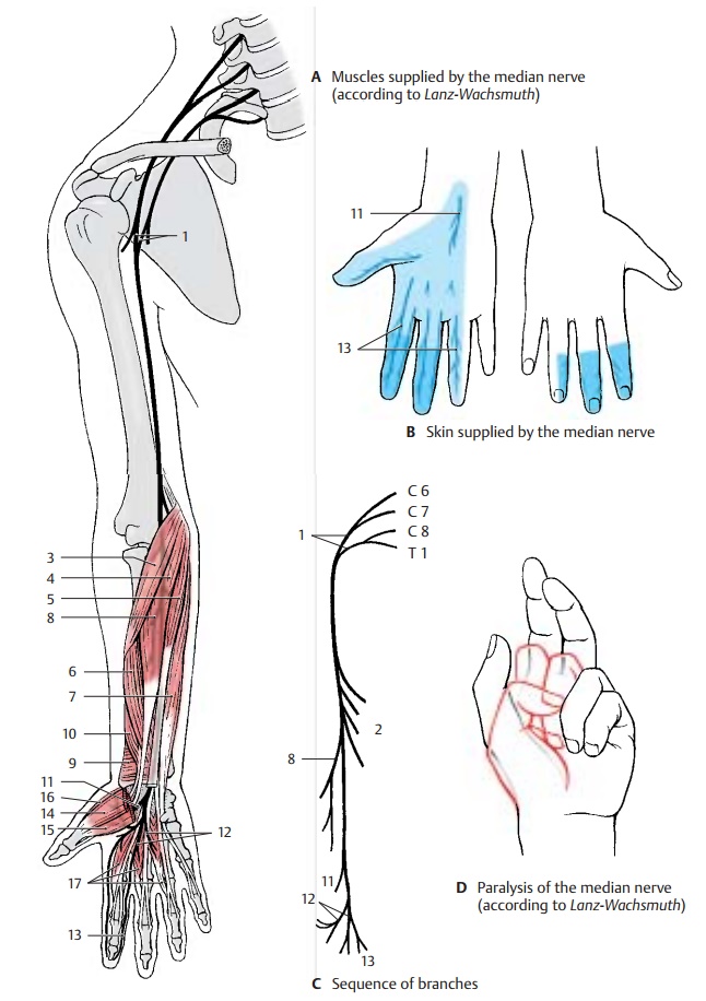
Median nerve (C6 – T1). Parts of the lateraland medial fascicles form the median loop (AC1) at the anterior surface of the axillary artery and join to form the median nerve.
The nerve extends in the medial bicipital sulcus along the surface of the brachial artery to the elbow, where it passes be-tween the two heads of the round pronator muscle to the forearm. It runs between the superficial flexor muscle of the fingers and the deep flexor muscle of the fingers to the wrist. Prior to its passage through the carpaltunnel, it lies superficially between the ten-dons of the radial flexor muscle of the wrist and the long palmar muscle. In the carpal tunnel, it ramifies into its terminal branches.
The muscular branches (C2) of the nerve supply the pronator muscles and most of the flexor muscles of the forearm, namely, theround pronator muscle (A3), the radial flexor muscle of the wrist (A4), the long pal-mar muscle (A5), and the superficial flexor muscle of the fingers with radial head (A6) and humeroulnar head (A7). In the elbow, the anterior interosseous nerve of the forearm (AC8) branches off and runs along the inter-osseous membrane to the quadrate prona-tor muscle (A9). It gives off branches to the long flexor muscle of the thumb (A10) and to the radial part of the deep flexor muscle of the fingers.
In the lower third of the forearm, the sensory palmar branch of the median nerve (A – C11) branches off to the skin of the ball of the thumb (thenar eminence), to the radial side of the wrist, and to the palm.
After passing through the carpal tunnel, the median nerve divides into three branches: the common palmar digital nerves I – III (A – C12), each of which bifurcates at the level of the metacarpophalangeal joints into two proper palmar digital nerves (A – C13). From the first common palmar digital nerve, a branch extends to the thenar eminence (short abductor muscle of thumb [A14], su-perficial head of the short flexor muscle of thumb [A15 ], and opposing muscle of thumb [A16]). The common palmar digitalnerves supply the lumbrical muscles I – III(A17). They run to the interdigits and bifur-cate in such a way that each pair of properpalmar digital nerves provides sensoryfibers to the lateral aspects of an interdigit. Thus, the first pair of nerves supplies the ulnar side of the thumb and the radial side of the index finger, the second pair supplies the ulnar side of the index finger and the radial side of the middle finger, and the third pair supplies the ulnar side of the middle finger and the radial side of the ring finger. The area innervated by the proper palmar digital nerves on the posterior side includes the end phalanx of the thumb as well as the distal and middle phalanges of the fingers (B).
The median nerve gives off branches to the periosteum, elbow joint, radiocarpal joint, and mediocarpal joint. At the level of the wrist, there is normally an anastomosis with the ulnar nerve.
Clinical Note: After injury to the nerve, pro-nation of the forearm is no longer possible and flexion is severely restricted. As to the hand, the thumb, index finger and middle finger can no longer be flexed at the end and middle phalanges, resulting in a characteristic feature of median pa-ralysis, the so-called hand of oath (D). On passing the carpal tunnel, the nerve can be injured by pressure in older persons (carpal tunnel syn-drome).
Innervation of the skin (B).Autonomiczone (dark blue) and maximum zone (light blue).
Related Topics