Chapter: Human Nervous System and Sensory Organs : The Ear
Inner Ear - Structure of The Ear
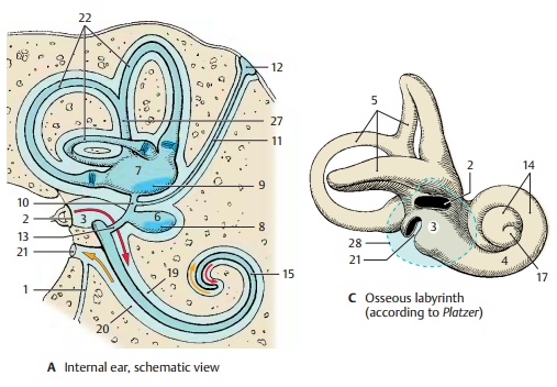
Inner Ear
The membranous labyrinth is a system of
vesicles and canals that is surrounded on all sides by a hard, bony capsule.
The cavities in the bone have the same shapes as the mem-branous structures, and
their cast (C) pro-vides a crude
representation of the mem-branous labyrinth. We therefore distinguish between
an osseous (bony) labyrinth and a membranous labyrinth. The osseous laby-rinthcontains a clear,
aqueous fluid, the perilymph (light
greenish-blue), in which themembranous labyrinth is suspended. The
perilymphatic space communicates with the subarachnoid space via the perilym-phatic duct (A1) at the posterior edge of thepetrous
bone. The membranous labyrinth contains the endolymph (dark greenish-blue) which is a viscous fluid.
The vestibular window (AC2) is closed by the stapes and leads into the middle part of the
osseous labyrinth, the vestibule of the
ear (AC3). The vestibule
communicates anteri-orly with the bony cochlea
(C4) and at its posterior wall with
the bony semicircularcanals (C5).

The vestibule contains two membranous
parts, the saccule (AB6) and the utricle (AB7). Both
structures contain sensory epithelium in a circumscribed part of the wall
(blue), the macula of the saccule (AB8) and the mac-ula of the utricle (AB9),
and are intercon-nected by the utriculosaccular
duct (AB10). The latter gives
off the slender endolym-phatic duct (A11) which runs to the poste-rior
surface of the petrous bone and ends beneath the dura mater as a flattened
ves-icle, the endolymphatic sac (A12). The unit-ing duct (AB13)
forms a connection betweenthe saccule and the membranous cochlear duct.
The osseous cochlea (C4) has about two and a half turns. The spiral canal of thecochlea (C14)
contains the membranous cochlear duct (AB15) which starts with ablind end, the
vestibular cecum (B16), and ends in the tip of the
cochlea, or cupula (C17), with the cupular cecum (B18). The
perilymphatic spaces are above and belowthe cochlear duct, or scala media; the scalavestibuli(AB19)
lies above it and opens intothe vestibule, and the scala tympani (AB20)
lies beneath it and is closed by the cochlearwindow
(A–C21).
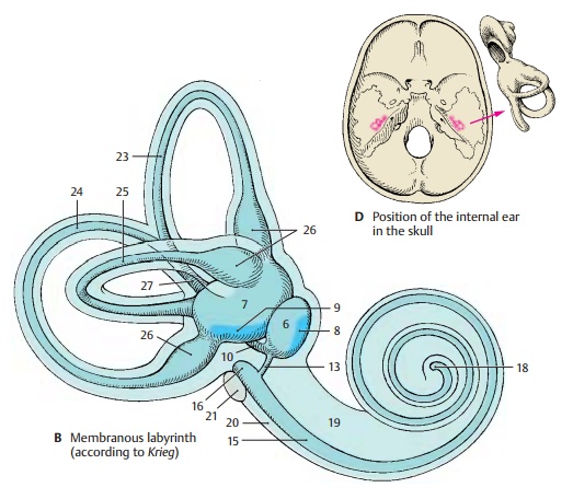
The
three bony semicircular canals (C5) emanating from the vestibule
contain the membranous semicircular
ducts (A22), which are connected
to the utricle. They are surrounded by perilymph and attached to the walls of
the perilymphatic space by con-nective-tissue fibers. The three semicircular
ducts are arranged perpendicularly to each other. The convexity of the anterior semi-circular duct (B23) is oriented toward thesurface of
the petrous pyramid, the posteriorsemicircular
duct (B24) runs parallel to
theposterior surface of the petrous bone, and the lateral semicircular duct (B25)
runs hori-zontally.
Each
semicircular duct has a dilatation at its transition to the utricle, the membranousampulla (B26), which corresponds to an os-seous ampulla in the bony canal.
The ante-rior and the posterior semicircular ducts join to form the common membranous crus(AB27). Each ampulla contains sensory
epithelium, the ampullary crest.
The
courses taken by the semicircular ducts do not correspond to the axes of the
body. The anterior and posterior semicircular ducts diverge from the median and
frontal planes by 45!; the lateral semicircular duct is
tilted in posterocaudal direction by 30!towards
the horizontal plane.
C28
Eardrum.
Related Topics