Chapter: Human Nervous System and Sensory Organs : The Eye
Eyelids - Structure of the Eye
Eyelids
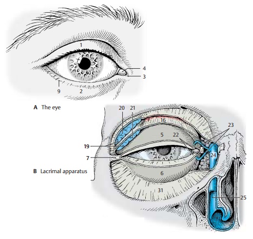
The eyeball is embedded in the orbit and is covered by the eyelids. The
upper eyelid (A1) and the lower eyelid
(A2) demarcate the palpebral fissure. The latter ends in
themedialangle of the eye (A3) with a recess enclosingthe lacrimal caruncle (A4).
In
Oriental People, the upper eyelid con-tinues medially onto the side of the nose
as a vertical fold of skin, the palpebronasal
fold. The fold can also be observed as a transitory formation in infants
and is known as epicanthus.

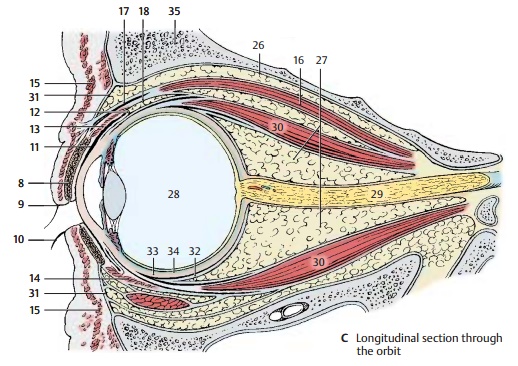
The
eyelids are reinforced by firm plates of connective tissue consisting of
collagenous fibers, tarsus superior (B5) and tarsus infe-rior(B6);
the tarsal plates, or palpebral car-tilages, are attached to the margin of the
orbit by the lateral palpebral ligament
(B7) and the medial palpebral ligament. The tar-sal plates contain elongated tarsal glands, the meibomian glands (C8), which spread over the entire
height of the eyelids. Their secretion prevents the tears from flowing over the
edge of the eyelids. They open at the posterior
margin of the eyelid. Several rows of eyelashes, the cilia (C10), emerge from
the anterior margin (AC9). The inner wall of the eyelids is
lined by the conjunctiva (C11), which extends to the anterior
aspect of the eyeball at the conjunctival
fornix (C12). The smooth superior tarsal muscle (C13) and inferior tarsal muscle (C14)
(inner-vated by the sympathetic nervous system), which control the size of the
palpebral fis-sure, attach at the tarsus. The eyelids are closed by the orbicular muscle of the eye(C15) (facial nerve, p. 122). The upper
lid is lifted by the levator muscle of
the upper eye-lid (BC16)
(oculomotor nerve), which at-taches at the upper margin of the optic canal. Its
superficial tendinous membrane (C17)
penetrates into the subcutaneous con-nective tissue of the upper lid, while the
deep tendinous membrane (C18)
attaches at the upper margin of the tarsus.
Related Topics