Chapter: Human Nervous System and Sensory Organs : Spinal Cord and Spinal Nerves
Cross Sections of the Spinal Cord
Cross Sections of the Spinal Cord
Cross
sections at different levels (left, myelin stain; right, cellular stain) vary
con-siderably. In the regions of cervical enlargement and lumbar enlargement,
the cross-sectional area is larger than in the rest of the spinal cord; it is
largest at the C4 – C5 and L4 – L5 levels. In both swellings, the numer-ous
nerves that supply the extremities cause an increase in gray matter.
The white matter is most extensive in the
cer-vical region and diminishes gradually in caudal direction; the ascending
sensory tracts increase in number from the sacral to the cervical region as
more fibers are added, while the descending motor tracts decrease from the
cervical to the sacral regions as fibers terminate at various levels.
The
butterfly configuration of the gray matter
changes in shape at the various levels, and so does the posterolateral tract (Lissauer’stract)
(A–D1).
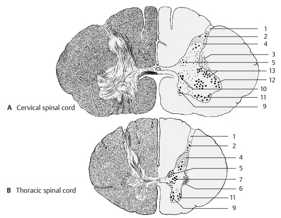
The posterior horn is narrow in the
cervical spinal cord; its tip ends in the cap-shaped marginal zone (nucleus posteromarginalis) (A2). The lateral angle between the
poste-rior and anterior horn is occupied by the re-ticular formation (AD3). The gelatinous sub-stance (Rolando’s substance) (A – D4) con-tains small, mostly peptidergic neurons where posterior
root fibers of various cali-bers terminate; it also contains descending fibers
from the brain stem (raphe nuclei, p. 108, B28; reticular formation, p. 146).
Un-myelinated processes of neurons ascend or descend for one to four root
levels within the posterolateral tract (Lissauer’s
tract) and then reenter into the gelatinous substance. Some of the
processes run within the lateral spinothalamic tract to the thalamus. The
fibers of proprioceptive sensi-bility in the muscles (muscle spindles)
ter-minate in the posterior thoracic
nucleus (dor-sal nucleus of Clarke)
(AB5) where the tractsto the
cerebellum begin. The reduced gray matter of the thoracic spinal cord has a
slender posterior horn with a prominent dorsal nucleus. In the plump posterior
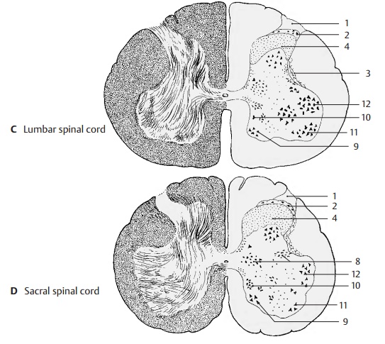
hornof the lumbar and sacral spinal cords, the gelatinous substance (CD4) is much en-larged and borders
dorsally on the narrow band of the marginal zone (CD2).

The lateral horn forms in the thoracic
spi-nal cord the lateral intermediate
substance (B6). It contains
sympathetic nerve fibers mainly for the vasomotor system, the effer-ent fibers
of which emerge via the anterior root. Sympathetic neurons also lie medially in
the intermediomedial nucleus (B7). In the sacral spinal cord,
parasympathetic neurons form the intermediolateral
nucleus und in-termediomedial nucleus
(D8).
The anterior horn expands in the cervical
spinal cord and contains several nuclei with large motor neurons, all of which
are cholin-ergic.
Medial group of nuclei
Anteromedial nucleus (A9)
Posteromedial nucleus (A10)
Lateral group of nuclei
Anterolateral nucleus (A11)
Posterolateral nucleus (A12)
Retroposterolateral nucleus (A13)
In the
region supplying the upper limbs, the anterior horn is far more differentiated
than in the thoracic spinal cord where only a few cell groups can be identified.
The expanded, plump anterior horn of the lumbar and sacral spinal cords, which
supplies the lower limbs, again contains several groups of nuclei.
Related Topics