Chapter: Medical Electronics : Bio-Chemical and Non Electrical Parameter Measurement
Blood Flow Meter
BLOOD FLOW METER
Blood
flow meters are used to monitor the blood flow in various blood vessels and to
measure cardiac output.
Types
·
Electromagnetic blood flow meters
·
Ultrasonic blood flow meters
·
Laser based blood flow meters
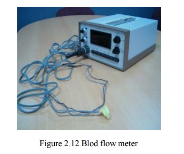
1. ELECTROMAGNETIC FLOWMETERS
§ Electromagnetic
blood flow meters measure blood flow in blood vessels
§ Consists
of a probe conneected to a flow sensor box
An
Electromagnetic Flow Met er is a device capable of measuring the mas s flow of
a fluid. Unlike the common flow meter you can find on the market it has no
moving parts, and for this reason it can be made to withstand any pressure
(without leakage)and any fluid(corrosive and non corrosive). This kind of flow
m eter use a magnet and two electrodes to p eek the voltage that appears across
the fluid moving in the magnetic field.

The Neumann Law (or Lenz Law) states that if a
conductive wire is moving at right angle through a magnetic field, a voltage E
[Volts] will appear at the end of the conductor (Fig.1):
E= B*L*V
Where
B =
Magnetic Induction
[Weber/m2]
L = Length of the portion of the wire 'wetted' by the
magnetic field
[m]
V =
Velocity of the wire [m/sec ]
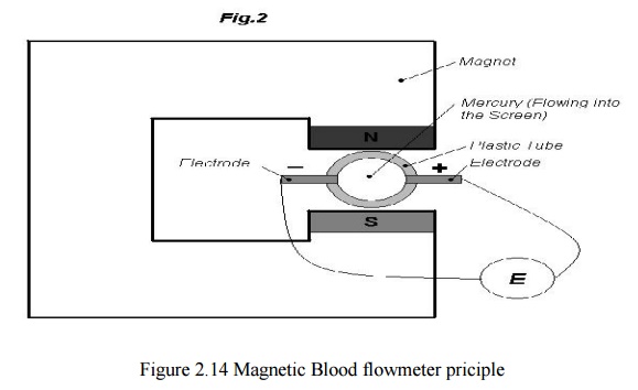
Now imagine you have a plastic tube with two
electrodes on the diameter and Mercury flowing into it (fig.2). A voltage will
appear on the electrodes and it will be
E=B*L*V
As in the
previous example (L in this case is the inner diameter of the tube).Mercury as
tiny conductive wires next to each other: each wire, moving in the tube, will
touch the two electrodes ,and thus you can mea sure their voltage.
An interesting
fact is t hat
if you reverse
the flow, you
still get a
voltage but with
reverse polarity (Fig.1). Till n
ow we have talked
about a conductive fluid ,Mercury, but this
stuff will also
work with non conductive fluid, provided that you use
an alternating magnetic field. Two
physicists, Middleman and Cushing, in an unpublished work, stated that when
using a non conductive f luid, if the frequency of the alternating magnetic
field is v the voltage at the electrodes will be attenuated by a factor a so
that:
Measuring the flow
`A perfect axisimmetri c construction cannot be
achieved and thus some magnetic flux lines will 'wet' t he connecting wires to
the electrodes. The alternating magnetic field will create an of fset voltage
in this wire and even if the fluid is not moving, the measured voltage will not
be zero.
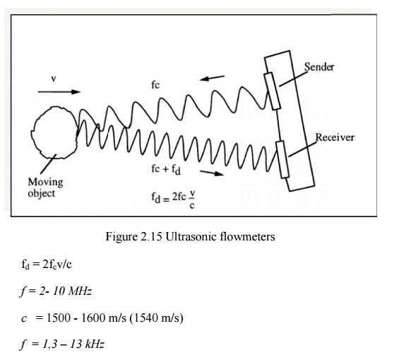
2. ULTRASONIC FLOWMETERS
The blood
cells in the fluid scatter the Doppler signal diffusively.In the recent years
ultrasound contrast agents have been used in order to increase the echoes.Th e
ultrasound beam is focused by a suitable transducer geometry and a lens.
In order
to know where a long the beam the blood flow data is colledted, a pulsed
Doppler must be used.The flow velocity is obtained from the spectral estimation
of the received Doppler signalThe ultrasound Doppler device can be either a continuous wave or a pulsed Doppler
A Continuous Wave
No
minimum range
Simpler
hardware
Range
ambiguity
Low flow
cannot be dete cted
A Pulsed Doppler
Accuracy
No
minimum flow
Minimum
range
(Maximum
flow) x (range)= limited the power decays exponentially because of the heating
of the tissue. The absorption coefficient ~ proportional to frequency the far
fi eld operation should be avoided due to beam divergence.

D = Transducer
diam eter (e.g. 1 – 5 mm) the backscattered power is proportional to f .The resolution and SNR are related to the pulse duration.
Improving either one of the parameters always affects inversely to the other
3. LASER DOPPLER FLO WMETRY
The
principle of measurement is the same as with ultrasound Doppler.The laser
parameter may have the following properties:5 mWH e-Ne-laser 632,8 nm
wavelength.
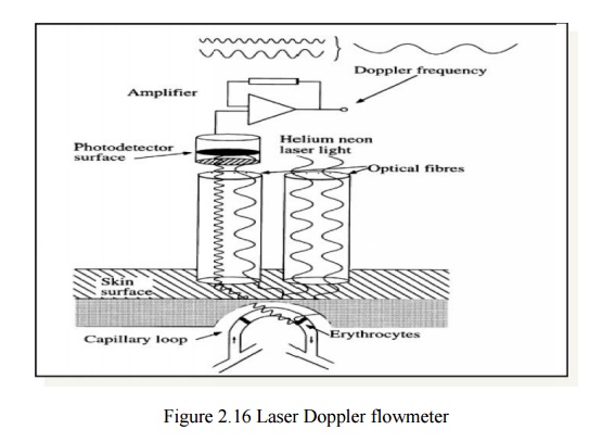
The
moving red blood cells cause Doppler frequency 30 – 12 0 00 Hz.The method is
used for capillary (microvascular) blood flow measurements
Indicator Dilution Methods
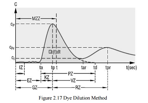
Dye Dilution Method
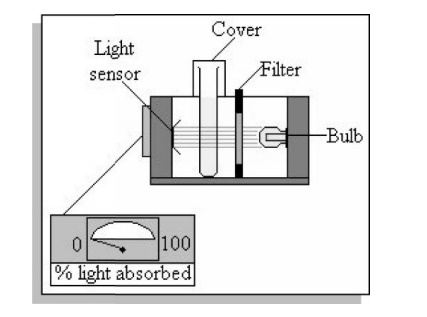
A bolus of indicator, a co lored dye (indocyanine green), is rapidly injected in to the vessel. The concentration is measured in the downstream The
blooddis drawn through a colorimetric cuvette and the concentration is
measured using the principle of absorption photometry
Thermal Dilution Method
A bolus
of chilled saline solution is injected into the blood circulation system
(right atrium). This causes decrease i n the pulmonary artery temperature. An
artery puncture is not needed in this technique.Several measurements can be done in relatively short time .A
standard technique for measuring cardiac output in critically ill patients
Photoelectric Method
A beam of
IR-light is directed to the part of the tissue which is to be measured for
blood flow (e.g. a finger or ear lobe)
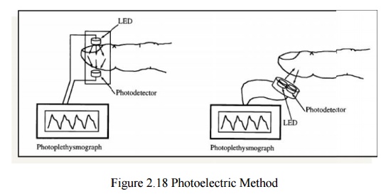
The blood
flow modulatees the attenuated / reflected light which is re corded.The light
that is transmitted / reflected is collected with a photodetector
Radioisotopes
A rapidly
diffusing, inert radioisotope of lipid-soluble gas ( Xe or Kr) is injected into
the tissue or passively diffused
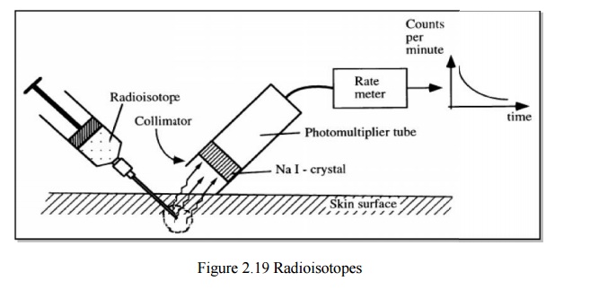
The
elimination of the radioisotope from microcirculatory bed is related to the
blood flow:
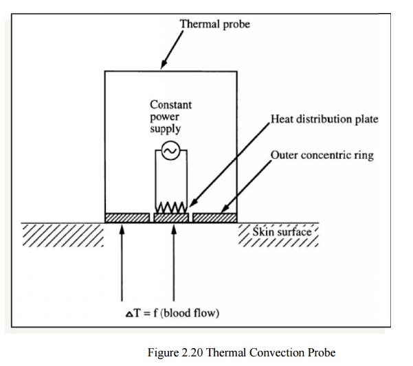
Thermal Convection Probe
·
This is one of the earliest techniques for blood
flow measurements
·
The rate of heat removal from the tissue under
probe is measured
·
The concentric rings are i solated thermally &
electrically from each other
The
central disk is heated 1 – 2 C over the temperature of tissue.A temperature
difference of 2- 3 C is established between the disks.The method is not very
common due extreme nonlinear properties and difficulties in practical use
(e.g. variable thermal characteristics of skin)
Related Topics