Chapter: 11th Zoology : Chapter 5 : Digestion and Absorption
Structure of the alimentary canal
Structure of
the alimentary canal
The
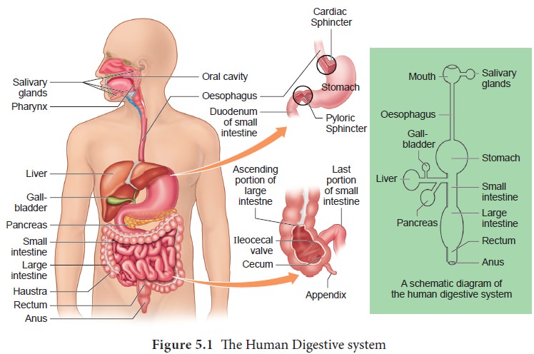
alimentary canal is a continuous, muscular digestive tract that begins with an
anterior opening, the mouth and opens out posteriorly through the anus. The
alimentary canal consists of mouth, buccal cavity, pharynx, oesophagus,
stomach, intestine, rectum and anus (Figure. 5.1). The mouth is concerned with
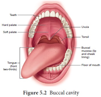
the reception of food and leads to the buccal cavity or oral cavity (Figure.
5.2). Mechanical digestion is initiated in the buccal cavity by chewing with
the help of teeth and tongue. Chemical digestion is through salivary enzymes
secreted by the salivary glands.
Each
tooth is embedded in a socket in the jaw bone; this type of attachment is
called thecodont. Human beings and
many mammals form two sets of teeth during their life time, a set of 20 temporary milk teeth (deciduous teeth) which
gets replaced by a set of 32 permanent teeth (adult teeth). This type of
dentition is called diphyodont. The permanent teeth are of four different types (heterodont),namely, Incisors- (I)
chisel like cutting teeth, -Canines (C) dagger shaped tearing teeth, Pre molars
(PM) for grinding, and Molars (M) for grinding and crushing. Arrangement of
teeth in each half of the upper and lower jaw, in the order of I, C, PM and M
can be represented by a dental formula, in human the dental- formula is
2123/2123.
Mineral
salts like calcium and magnesium are deposited on the teeth and form a hard
layer of ‘tartar’ or calculus called
plaque. If the plaque formed on teeth is not removed regularly, it would spread
down the tooth into the narrow gap between- the gums and enamel and causes
inflammation, called gingivitis,
which leads to redness and bleeding of the gums and to bad smell. The hard
chewing surface of the teeth is made of enamel and helps in mastication of
food.
Tongue is a freely movable muscular organ attached at the posterior end by the frenulum to the floor of the buccal cavity and is free in the front. It acts as a universal tooth brush and helps in intake food, chew and mix food with saliva, to swallow food and also to speak. The upper surface of the tongue has small projections called papillae with taste buds.
The oral
cavity leads into a short common passage for food and air called pharynx. The
oesophagus and the trachea (wind pipe) open into the pharynx. Food passes into
the oesophagus through a wide opening called gullet at the back of the pharynx.
A cartilaginous flap called -epiglottis prevents the entry of food into the
glottis (opening of trachea) during swallowing. Two masses of lymphoid tissue-
called tonsils are also located at the sides of the pharynx.
Oesophagus
is a thin long muscular tube concerned with conduction of the food to a ‘J’
shaped stomach passing through the neck, thorax and diaphragm. A cardiac
sphincter (gastro oesphageal sphincter) regulates the opening of oesophagus
into the stomach (Figure. 5.1). If the cardiac sphincter does not contract
properly during the churning action of the stomach the gastric juice with acid
may flow back into the oesophagus and cause heart burn, resulting in GERD (Gastero Oesophagus Reflex
Disorder).
The
stomach functions as the temporary storage organ for food and is located in the
upper left portion of the abdominal cavity. It consists of three parts – a
cardiac portion into which the oesophagus opens; a fundic portion and a pyloric
portion that opens into the duodenum. The opening of the stomach into the
duodenum is guarded by the pyloric sphincter. It periodically allows partially
digested food to enter the duodenum and also prevents
regurgitation of food. The inner wall of stomach has many folds called gastric rugae which unfolds to
accommodate a large meal.
The small
intestine assists in the final digestion and absorption of food. It is the
longest part of the alimentary canal and has three regions, a ‘U’ shaped
duodenum (25cm long), a long coiled middle portion jejunum (2.4m long) and a
highly coiled ileum (3.5m long). The wall of the duodenum has Brunner’s glands
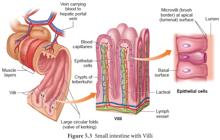
which secrete mucus and enzymes. Ileum is the longest part of the small
intestine and opens into the caecum of the large intestine. The ileal mucosa
has numerous vascular projections called villi which are involved in the
process of absorption and the cells lining the villi produce numerous
microscopic projections called microvilli giving a brush border appearance that
increase the surface area enormously. Along with villi, the ileal mucosa also
contain mucus secreting goblet cells and lymphoid tissue known as Peyer’s patches which produce
lymphocytes. The wall of the small intestine bears crypts between the base of villi called crypts of Leiberkuhn(Figure.5.3).
The large
intestine consists of caecum, colon and rectum. The caecum is a small blind
pouch like structure that opens into the colon and it possesses a narrow finger
like tubular projection called vermiform
appendix . Both caecum and vermiform appendix are large in herbivorous
animal and act as an important site for cellulose
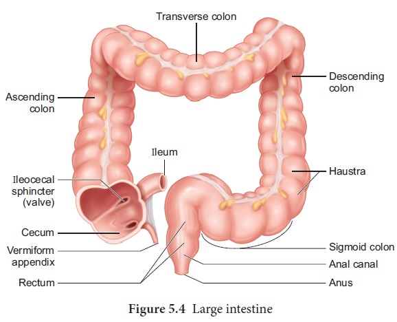
The colon is divided into four regions –
an ascending, a transverse, a descending part and a sigmoid colon. The colon is
lined by dilations called haustra (singular
– haustrum) (Figure.5.4). The “S” shaped sigmoid colon (pelvic colon) opens
into the rectum. Rectum is concerned with temporary storage of faeces. The
rectum open out through the anus. The anus is guarded by two anal sphincter
muscles. The anal mucosa is folded into several vertical folds and contains
arteries and veins called anal columns. Anal column may get enlarged and causes
piles or haemorrhoids.
Related Topics