Structure, Process - Human Digestive system | 11th Zoology : Chapter 5 : Digestion and Absorption
Chapter: 11th Zoology : Chapter 5 : Digestion and Absorption
Human Digestive system
Digestive
system
The
process of digestion involves intake of the food (Ingestion), breakdown of the
food into micromolecules (Digestion), absorption of these molecules into the
blood stream (Absorption), the absorbed substances becoming components of cells
(Assimilation) and elimination of the undigested substances (Egestion).
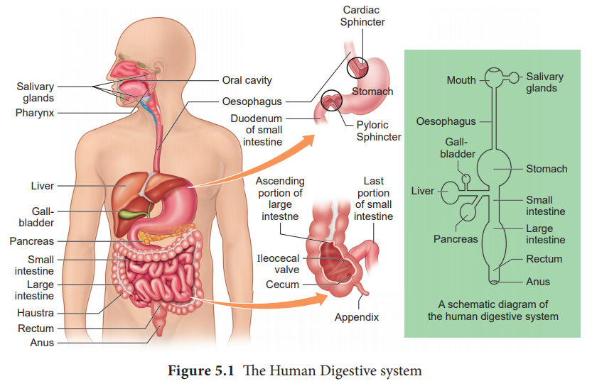
Digestive system includes the alimentary canal and associated digestive glands.
1. Structure of the alimentary canal
The
alimentary canal is a continuous, muscular digestive tract that begins with an
anterior opening, the mouth and opens out posteriorly through the anus. The
alimentary canal consists of mouth, buccal cavity, pharynx, oesophagus,
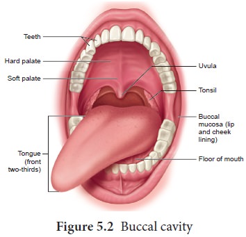
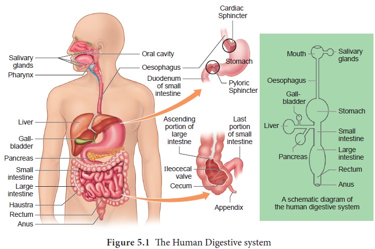
stomach, intestine, rectum and anus (Figure. 5.1). The mouth is concerned with
the reception of food and leads to the buccal cavity or oral cavity (Figure.
5.2). Mechanical digestion is initiated in the buccal cavity by chewing with
the help of teeth and tongue. Chemical digestion is through salivary enzymes
secreted by the salivary glands.
Each
tooth is embedded in a socket in the jaw bone; this type of attachment is
called thecodont. Human beings and
many mammals form two sets of teeth during their life time, a set of 20 temporary milk teeth (deciduous teeth) which
gets replaced by a set of 32 permanent teeth (adult teeth). This type of
dentition is called diphyodont. The permanent teeth are of four different types (heterodont),namely, Incisors- (I)
chisel like cutting teeth, -Canines (C) dagger shaped tearing teeth, Pre molars
(PM) for grinding, and Molars (M) for grinding and crushing. Arrangement of
teeth in each half of the upper and lower jaw, in the order of I, C, PM and M
can be represented by a dental formula, in human the dental- formula is
2123/2123.
Mineral
salts like calcium and magnesium are deposited on the teeth and form a hard
layer of ‘tartar’ or calculus called
plaque. If the plaque formed on teeth is not removed regularly, it would spread
down the tooth into the narrow gap between- the gums and enamel and causes
inflammation, called gingivitis,
which leads to redness and bleeding of the gums and to bad smell. The hard
chewing surface of the teeth is made of enamel and helps in mastication of
food.
Tongue is a freely movable muscular organ attached at the posterior end by the frenulum to the floor of the buccal cavity and is free in the front. It acts as a universal tooth brush and helps in intake food, chew and mix food with saliva, to swallow food and also to speak. The upper surface of the tongue has small projections called papillae with taste buds.
The oral
cavity leads into a short common passage for food and air called pharynx. The
oesophagus and the trachea (wind pipe) open into the pharynx. Food passes into
the oesophagus through a wide opening called gullet at the back of the pharynx.
A cartilaginous flap called -epiglottis prevents the entry of food into the
glottis (opening of trachea) during swallowing. Two masses of lymphoid tissue-
called tonsils are also located at the sides of the pharynx.
Oesophagus
is a thin long muscular tube concerned with conduction of the food to a ‘J’
shaped stomach passing through the neck, thorax and diaphragm. A cardiac
sphincter (gastro oesphageal sphincter) regulates the opening of oesophagus
into the stomach (Figure. 5.1). If the cardiac sphincter does not contract
properly during the churning action of the stomach the gastric juice with acid
may flow back into the oesophagus and cause heart burn, resulting in GERD (Gastero Oesophagus Reflex
Disorder).
The
stomach functions as the temporary storage organ for food and is located in the
upper left portion of the abdominal cavity. It consists of three parts – a
cardiac portion into which the oesophagus opens; a fundic portion and a pyloric
portion that opens into the duodenum. The opening of the stomach into the
duodenum is guarded by the pyloric sphincter. It periodically allows partially
digested food to enter the duodenum and also prevents
regurgitation of food. The inner wall of stomach has many folds called gastric rugae which unfolds to
accommodate a large meal.
The small
intestine assists in the final digestion and absorption of food. It is the
longest part of the alimentary canal and has three regions, a ‘U’ shaped
duodenum (25cm long), a long coiled middle portion jejunum (2.4m long) and a
highly coiled ileum (3.5m long). The wall of the duodenum has Brunner’s glands
which secrete mucus and enzymes. Ileum is the longest part of the small
intestine and opens into the caecum of the large intestine. The ileal mucosa
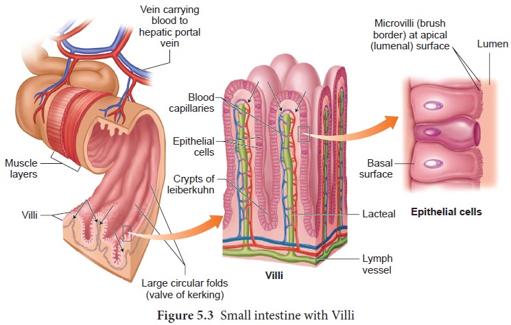
has numerous vascular projections called villi which are involved in the
process of absorption and the cells lining the villi produce numerous
microscopic projections called microvilli giving a brush border appearance that
increase the surface area enormously. Along with villi, the ileal mucosa also
contain mucus secreting goblet cells and lymphoid tissue known as Peyer’s patches which produce
lymphocytes. The wall of the small intestine bears crypts between the base of villi called crypts of Leiberkuhn(Figure.5.3).
The large
intestine consists of caecum, colon and rectum. The caecum is a small blind
pouch like structure that opens into the colon and it possesses a narrow finger
like tubular projection called vermiform
appendix . Both caecum and vermiform appendix are large in herbivorous
animal and act as an important site for cellulose
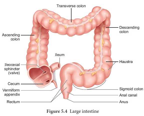
The colon is divided into four regions –
an ascending, a transverse, a descending part and a sigmoid colon. The colon is
lined by dilations called haustra (singular
– haustrum) (Figure.5.4). The “S” shaped sigmoid colon (pelvic colon) opens
into the rectum. Rectum is concerned with temporary storage of faeces. The
rectum open out through the anus. The anus is guarded by two anal sphincter
muscles. The anal mucosa is folded into several vertical folds and contains
arteries and veins called anal columns. Anal column may get enlarged and causes
piles or haemorrhoids.
2. Histology of the Gut
The wall
of the alimentary canal from oesophagus to rectum consists of four layers
(Figure 5.5) namely serosa, muscularis, sub-mucosa and mucosa.
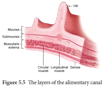
The serosa (visceral peritoneum) is the
outermost layer and is made up of thin squamous epithelium with some connective
tissues. Muscularis is made of smooth circular and longitudinal muscle fibres
with a network of nerve cells and parasympathetic nerve fibres which controls
peristalsis. The -submucosal layer is formed of loose connective tissue
containing nerves, blood, lymph vessels and the sympathetic nerve fibres that
control the secretions of intestinal juice.
The
innermost layer lining the lumen of the alimentary canal is the mucosa which
secretes mucous.
3. Digestive glands
Digestive
glands are exocrine glands which secrete biological catalysts called enzymes.
The digestive glands associated with the alimentary canal are salivary glands,
liver and pancreas. Stomach wall has gastric glands that secrete gastric juice
and the intestinal mucosa secretes intestinal juice.
Salivary glands
There are
three pairs of salivary glands in the mouth. They are the largest parotids
gland in the cheeks, the sub-maxillary/ sub-mandibular in the lower jaw and the
sublingual beneath the tongue. These glands have ducts such as Stenson’s duct,
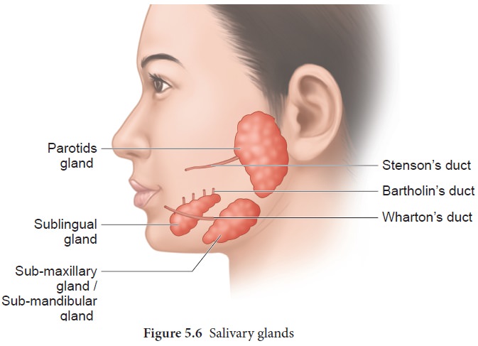
Wharton’s duct and Bartholin’s duct or duct of
Rivinis respectively (Figure. 5.6). The
salivary juice secreated by the salivary glands reaches the mouth through these
ducts. The daily secretion of saliva from salivary glands ranges from 1000 to
1500mL
Gastric glands
The wall
of the stomach is lined by gastric glands. Chief cells or peptic cells or zymogen
cells in the gastric glands secrete gastric enzymes and Goblet cells secrete mucus.
The Parietal or oxyntic cells
secrete HCl and an intrinsic factor responsible for the absorption of Vitamin B12
called Castle’s intrinsic factor.
Liver
The
liver, the largest gland in our body is situated in the upper right side of the
abdominal cavity, just below the diaphragm. The liver consists of two major
left and right lobes; and two minor lobes. These lobes are connected with
diaphragm. Each lobe has many hepatic lobules (functional unit of liver) and is
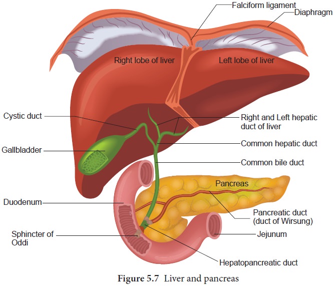
covered by a thin connective tissue sheath called the Glisson’s capsule. Liver cells (hepatic cells) secrete
bile which is stored and concentrated in a thin muscular sac called the gall
bladder. The duct of gall bladder (cystic duct) along with the hepatic duct
from the liver forms the common bile duct. The bile duct passes downwards and
joins with the main pancreatic duct to form a common duct called
hepato-pancreatic duct. The opening of the hepato-pancreatic duct into the
duodenum is guarded by a sphincter called the sphincter of Oddi (Figure.5.7).
Liver has high power of regeneration and liver cells are replaced by new ones
every 3-4 weeks.
Apart
from bile secretion, the liver also performs several functions
1. Destroys
aging and defective blood cells
2. Stores
glucose in the form of glycogen or disperses glucose into the blood stream with
the help of pancreatic hormones
3. Stores
fat soluble vitamins and iron
4. Detoxifies
toxic substances.
5. Involves
in the synthesis of non-essential amino acids and urea.
Pancreas
The
second largest gland in the digestive system is the Pancreas, which is a yellow
coloured, compound elongated organ consisting of exocrine and endocrine cells.
It is situated between the limbs of the ‘U’ shaped duodenum. The exocrine
portion secretes pancreatic juice containing enzymes such as pancreatic amylase,
trypsin and pancreatic lipase and the endocrine part called Islets of
Langerhans secretes hormones such as insulin and glucagon. The pancreatic duct
directly opens into the duodenum.
Related Topics