Chapter: The Massage Connection ANATOMY AND PHYSIOLOGY : Muscular System
Structure of Skeletal Muscle
STRUCTURE OF SKELETAL MUSCLE
Macroscopic Structure of Muscle
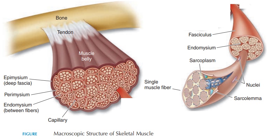
Skeletal muscle, as we know it (e.g., biceps brachii), is a collection of muscle cells, nerves, connective tis-sue, and blood vessels. Each muscle cell is known as a muscle fiber. Muscle fibers are cylindrical, arranged parallel to each other, and run through the entire length of the muscle. The fibers are held in place by connective tissue, which surround individ-ual fibers, bundles of muscle fibers and, finally, the entire muscle. It is the connective tissue that attaches muscle to the periosteum of bones and conveys the force generated by the muscle to the bone, across one or more joints. Other than the connective tissue sur-rounding individual fibers, bundles of fibers, and the entire muscle, connective tissue (fascia) separates muscle from the skin (superficial fascia or subcuta-neous layer) and holds groups of muscles with simi-lar functions together (deep fascia).
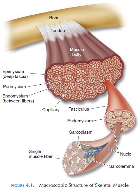
The connective tissue that surrounds an individual fiber is the endomysium (see Figure 4.1). Fascicles are bundles of muscle fibers surrounded by addi-tional connective tissue, the perimysium. The per-imysium attaches adjacent fascicles together in addi-tion to carrying blood vessels and nerves to the muscle fibers. The whole muscle is surrounded by connective tissue called the epimysium. This con-nective tissue (part of the deep fascia) separates mus-cles from each other and the surrounding organs. The epimysium, in turn, is continuous, with a rope-like connective tissue—the tendon or connective tis-sue sheet—aponeurosis. The tendon or aponeurosis ultimately weaves intimately with the periosteum of bone, attaching the muscle. By this interconnection of connective tissue, the power generated by the con-traction of individual muscle fibers is conveyed to the bone. The fleshy part of the muscle that lies between the connective tissue that attaches it to both ends of the bone is known as the belly of the muscle.
Microscopic Structure of Individual Muscle Fibers
Knowledge of the microscopic structure (see Figure 4.2) is required to understand the contractile process of muscle. The shape of the muscle fiber is cylindrical, and each fiber extends through the length of the muscle.
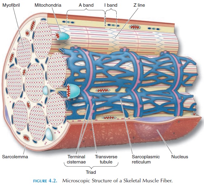
Each fiber appears striated under a microscope (i.e., having dark and light bands); it is referred to as striated muscle (the cause of the striated appear-ance is explained later). The muscle fiber, like all other cells, has many cell organelles. The cytoplasm, known here as sarcoplasm, is enclosed in a cell membrane called the sarcolemma.The skeletal muscle fiber is multinucleated and has hundreds of nuclei located just below the cell membrane. It is be-lieved that the number of nuclei denotes the number of embryonic muscle cells (myoblasts) that have fused to form one cell fiber. Certain myoblasts (satel-lite cells) do not fuse and are seen as individual cellsbetween the muscle fibers. The satellite cells may en-large, divide, and fuse with damaged muscle fibers, assisting in the regeneration of tissue. However, re-generation of muscle tissue is minimal and cells can-not divide.
Muscle fiber has numerous tubes, known as Ttubules or transverse tubules, that run transversely into the sarcoplasm. The tubules are continuous with the sarcolemma and transmit impulses generated by a nerve into the cell. Inside the sarcoplasm, the T tubules encircle the myofibrils, long cylindrical structures that extend the entire length of the muscle fiber. Hundreds of myofibrils are seen in each muscle fiber. Each myo-fibril is actually a collection of specialized proteins called myofilaments. The activity of the myofilaments produce contraction and relaxation of the muscle.
Other than the transverse tubules, which are actu-ally invaginations of the sarcolemma into the sar- coplasm, there is another network of tubules (sar-coplasmic reticulum[SR]) in the sarcoplasm. SR isequivalent to the endoplasmic reticulum of other cells; it surrounds individual myofibrils on each side of the T tubules. Close to the T tubules, SR is enlarged to form an expanded chamber called the terminalcisternae. SR contains a high concentration of cal-cium ions that is required for muscle contraction.
Sarcoplasm also contains glycogen—storage forms of glucose that can be broken down during metabo-lism. In addition, sarcoplasm contains a red, hemo-globinlike protein called myoglobin. Myoglobin, similar to hemoglobin, is capable of binding oxygen. This oxygen is used by the mitochondria for adeno-sine triphosphate (ATP) production.
Related Topics