Chapter: The Massage Connection ANATOMY AND PHYSIOLOGY : Muscular System
Cardiac, Smooth, and Skeletal Muscle

Cardiac, Smooth, and Skeletal Muscle
The basic contractile process is the same in cardiac, smooth, and skeletal muscle, with movement produced by the action of the myofilaments actin and myosin. However, because the requirements in terms of speed and force of contraction are different, the structure of cardiac and smooth are slightly different than skeletal muscle.
CARDIAC MUSCLE
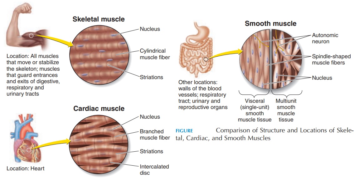
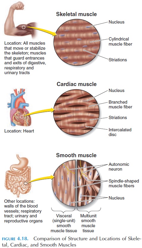
The cardiac muscle (see Figure 4.18;) present in the walls of the heart is used to propel blood from the chambers, requiring each chamber to contract in one accord. Relaxation should also be synchronous for blood to fill the chamber. To meet these needs, the structure is altered.

Cardiac muscle is branched and has specialized re-gions on the sarcolemma where it comes in contact with the adjoining cell. These specialized regions, in-tercalated disks, contain proteins (desmosomes) that hold adjacent cells together and transmit the force generated from muscle to muscle. Intercalated disks also contain gap junctions, which are specialized channels that allow action potentials (impulses) to travel from one cell to another. Because of the pres-ence of intercalated disks, cardiac muscle is able to contract together as a functional syncytium (as if it functioned as one muscle fiber). The myosin and actin filaments are arranged in an orderly manner; and cardiac muscle, like skeletal muscle, looks stri-ated.
Because the heart must alter its force of contrac-tion according to regional requirements, its contrac-tion is not only regulated by nerves, but also by hor-mones and ionic contents of the blood among others. For example, adrenaline in blood can speed contrac-tion and calcium levels can alter the excitability and contractility of the heart. Unlike skeletal muscle that relies on a stimulus from a nerve fiber, the cardiac muscle can respond to action potentials produced by specialized cardiac muscle fibers belonging to the conducting system of the heart. In re-sponse to a stimulus, the cardiac muscle fiber re-mains contracted for a longer period (about 10 to 15 times the duration of skeletal muscle).
Cardiac muscle does not have the capacity to re-generate.
SMOOTH MUSCLE
Smooth muscle is spindle-shaped with no striations. Because the demands made of smooth muscle for speed and force of contraction is considerably less, actin and myosin filaments are scattered in the cyto-plasm; hence, the lack of striations. The sarcoplasm does not contain transverse tubules and has few sar-coplasmic reticulum for calcium storage. However, smooth muscle has specialized calcium binding reg-ulatory proteins called calmodulin, which is similar to troponin in skeletal muscle. The sarcoplasm of smooth muscle contains scattered filaments (densebodies) that are equivalent to the Z disks in the skele-tal muscle. Dense bodies are also found attached to the sarcolemma. Other filaments interconnect adja-cent dense bodies. The force generated by the sliding between the scattered actin and myosin filaments is generated to the dense bodies that, in turn, cause shortening of the muscle fiber.
Smooth muscle contractions are slower and last longer than those of skeletal or cardiac muscle. The lack of T tubules and scarce sarcoplasmic reticulum for calcium is one reason for the slower start and longer duration; therefore, it takes longer for calcium to diffuse into the cell and initiate sliding between the actin and myosin. The shortening produced in smooth muscle is also considerable, a result of the slow diffusion of calcium from the cell to the outside.
A special feature of smooth muscle is the ability to stretch and shorten to a greater extent and still main-tain contractile function. Smooth muscle, like skele-tal muscle, has muscle tone. This is important in the gut where the walls must maintain a steady pressure on the contents. It is also important in blood vessels that must maintain pressure.
Smooth muscle, similar to cardiac muscle, re-sponds to changes in the local environment and fac-tors such as hormones, ions, pH, temperature, and stretch.
There are two types of smooth muscle. The singleunit, or visceral smooth muscle fibers, form largenetworks and are connected by gap junctions. This enables the smooth muscle to contract in waves when stimulated at one end of the organ. This is the more common type and it is found in the walls of small ar-teries and veins and walls of hollow organs.
The multiunit smooth muscle fibers act indepen-dently. Similar to skeletal muscle fiber, each muscle fiber is innervated, with few gap junctions between adjacent cells. Therefore, each fiber must be stimu-lated separately to produce contraction. These fibers are found in the walls of large arteries, bronchioles, arrector pili muscle attached to hair follicles, and muscles of the eye that control the size of the pupil and shape of the lens.
Smooth muscle can regenerate more than other muscle tissue, but much less than epithelial tissue.
Related Topics