Chapter: The Massage Connection ANATOMY AND PHYSIOLOGY : Muscular System
Appendicular Musculature - Origin and Insertion of Muscles
THE APPENDICULAR MUSCULATURE
The appendicular musculature includes muscles that help stabilize and position the pectoral and pelvic gir-dle and move the upper and lower limbs.
To make it more practical and applicable to body-workers, the muscles of the pectoral girdle and upper limbs are described in four groups; each group, in turn, being subdivided according to the movements they perform. The four groups are: (1) Muscles that position and move the shoulder girdle; (2) Muscles that move the arm; (3) Muscles that move the forearm and wrist; and (4) Muscles that move the palm and fingers.
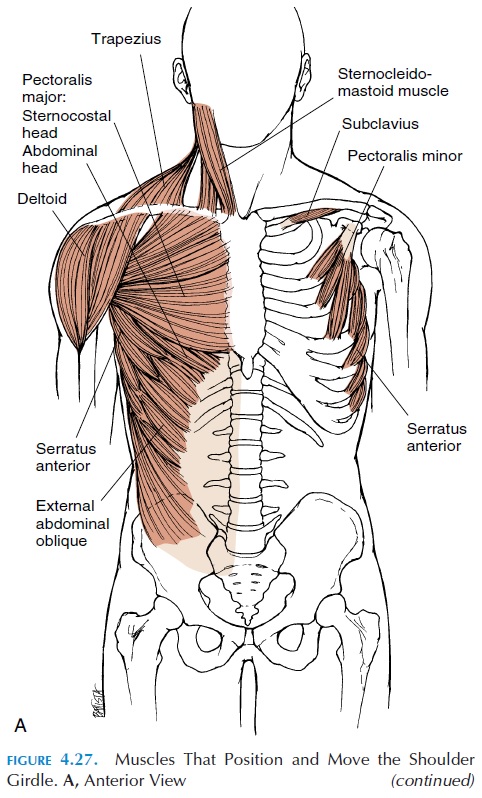
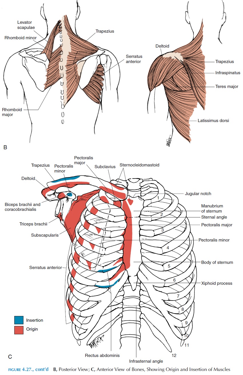
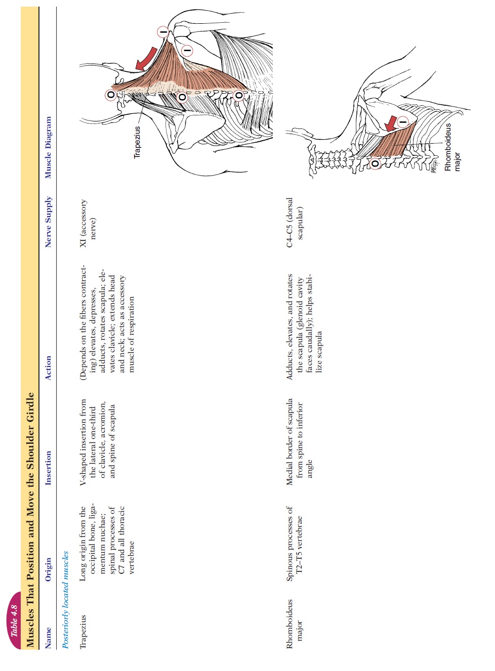
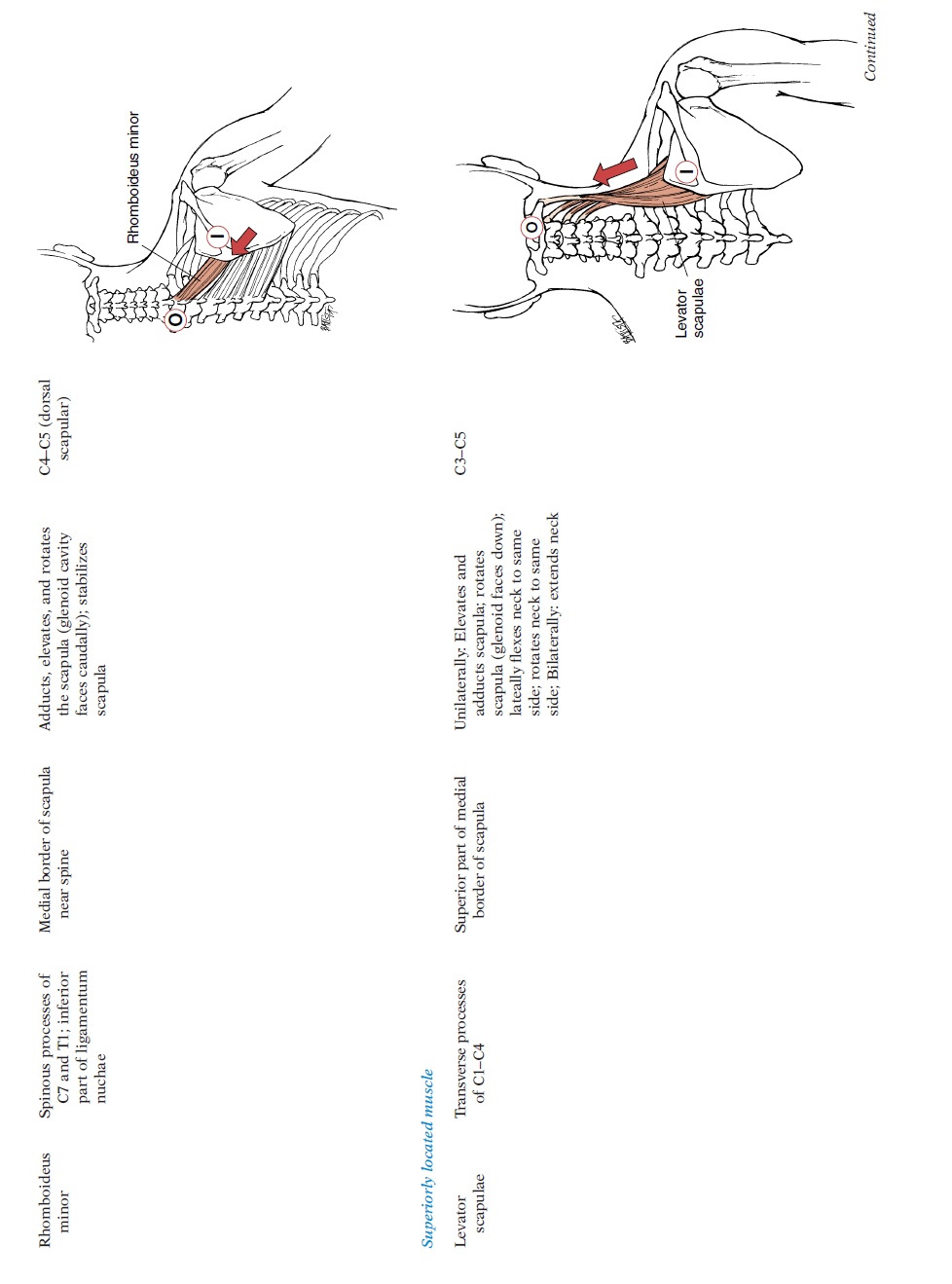
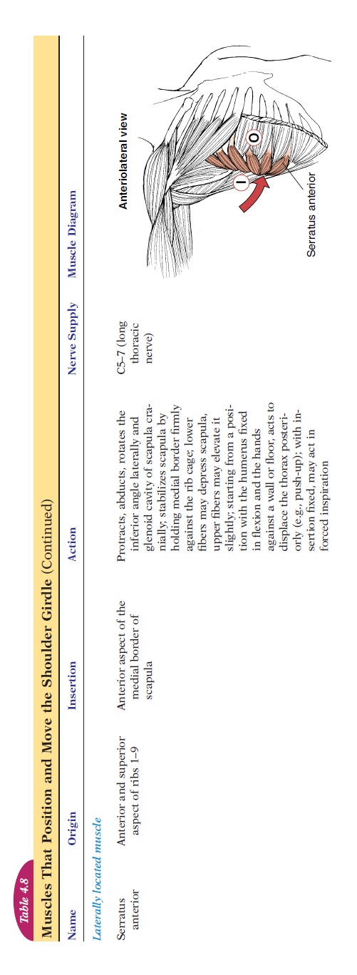
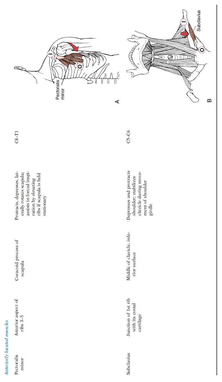
Muscles That Position andMove the Shoulder Girdle
These muscles (see Figure 4.27 Table 4.8) stabilize the scapula and clavicle. As mentioned, the only joint in the pectoral girdle that articulates with the axial skeleton is at the stern-oclavicular joint. The scapula and the clavicle are held in place by the numerous muscles that originate from the axial skeleton.
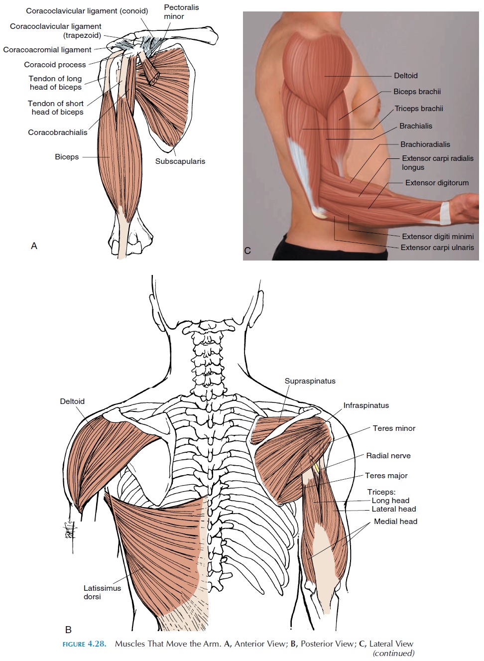
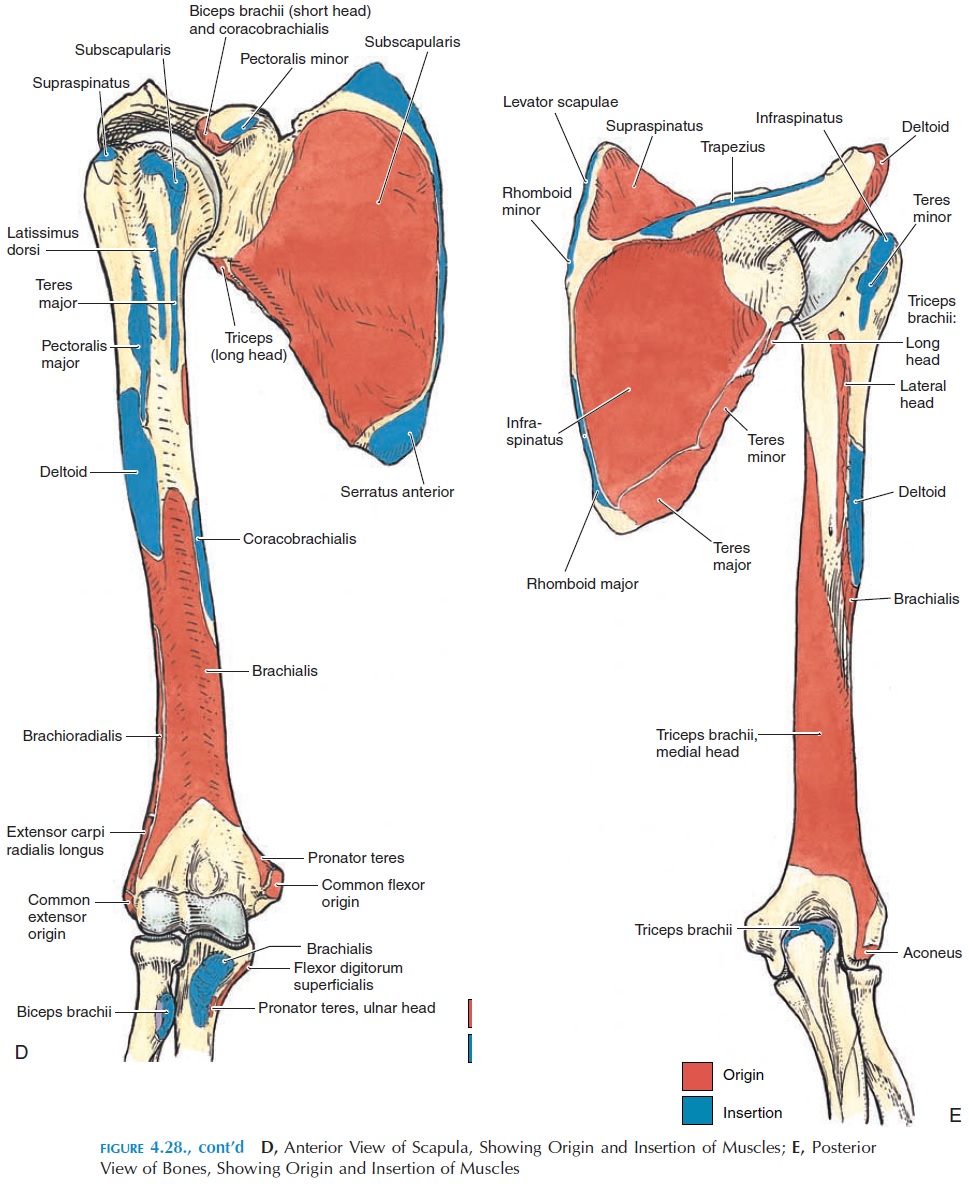
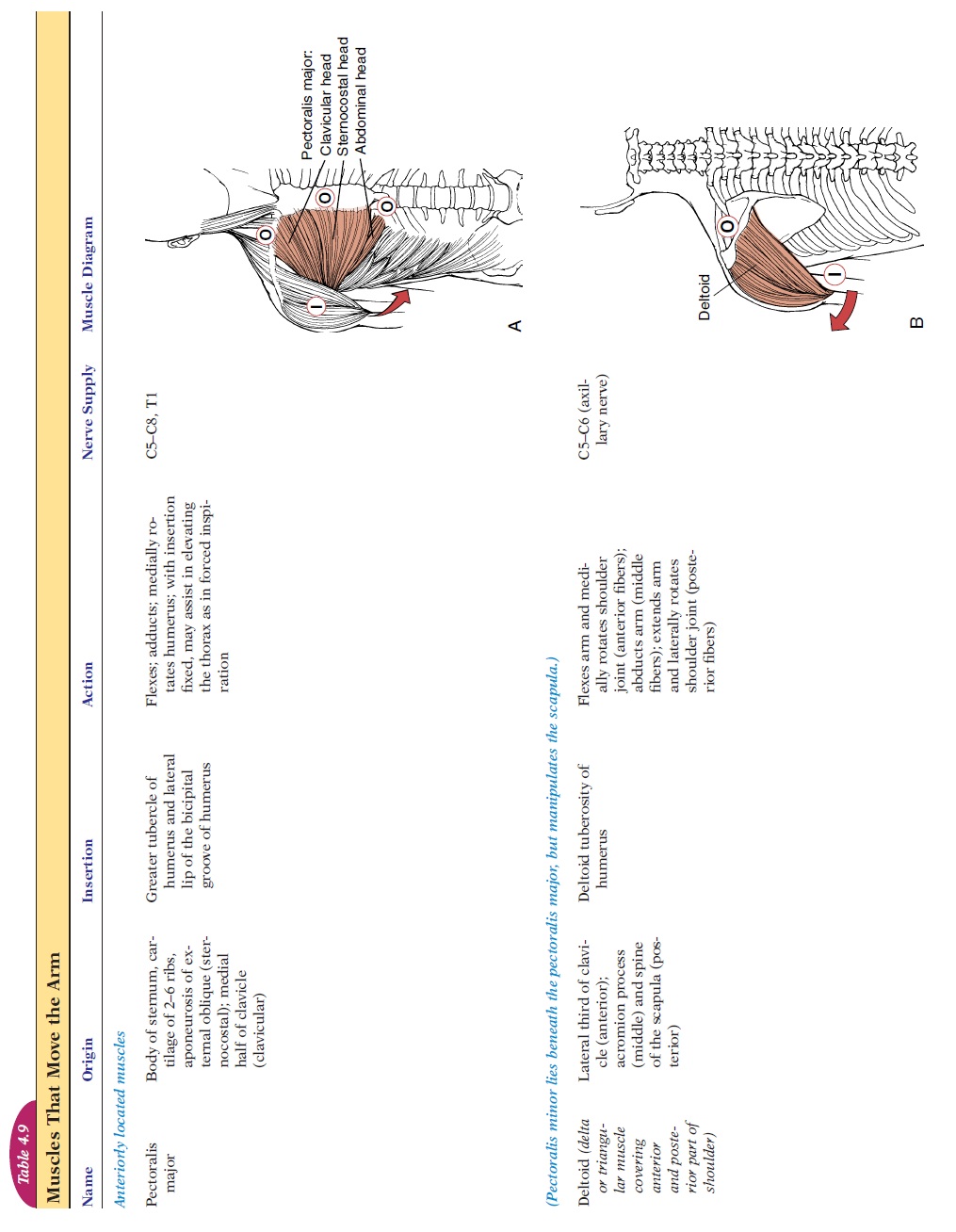
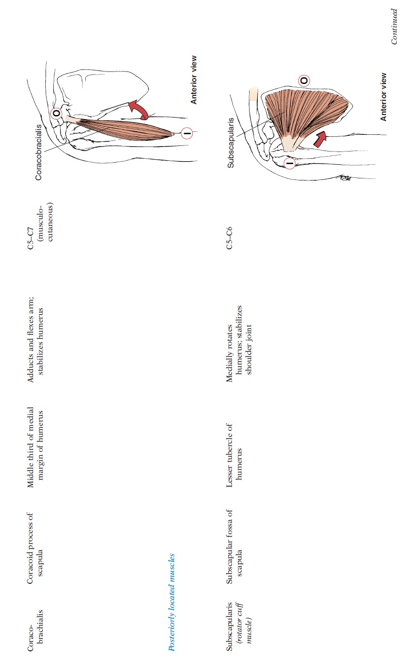
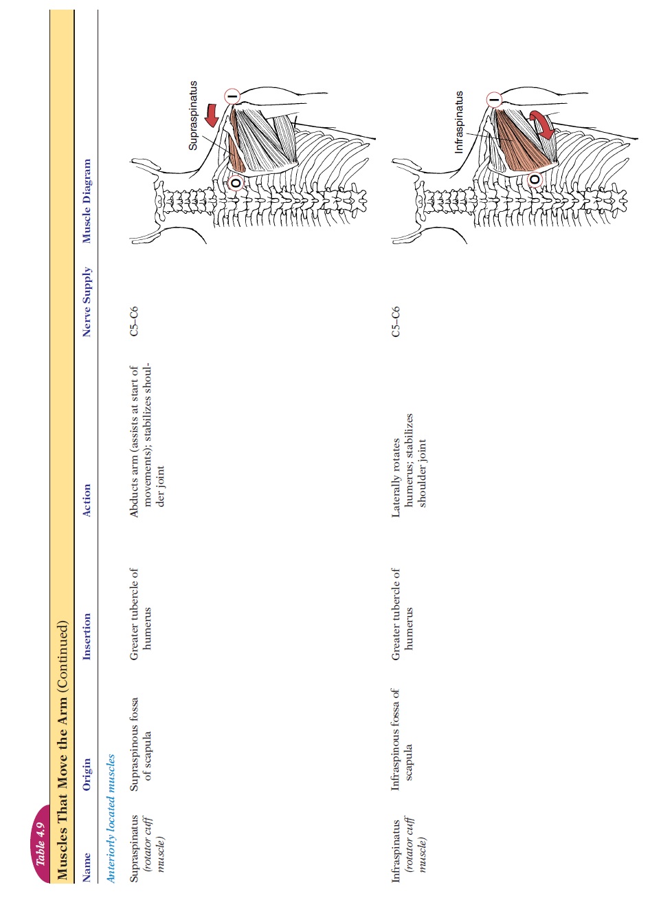
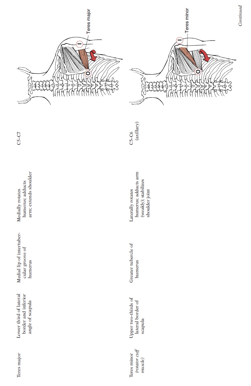
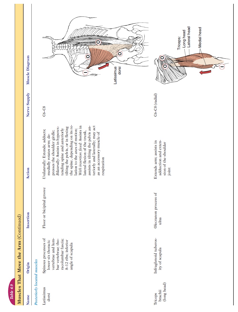
Muscles That Move the Arm
The muscles that move the arm (see Figure 4.28 , Table 4.9) cross the shoulder joint and attach to the humerus, around or close to the humeral head. They originate posteriorly from the scapula and the vertebrae. Anteriorly, the muscles originate from the sternum, the cartilage of ribs 2–6, and the clavicle.
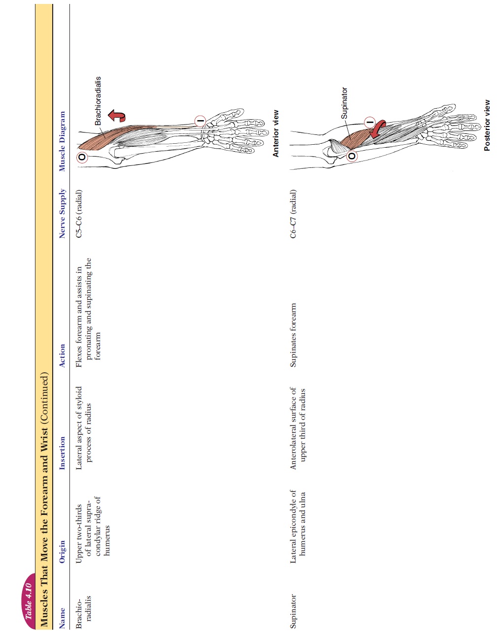
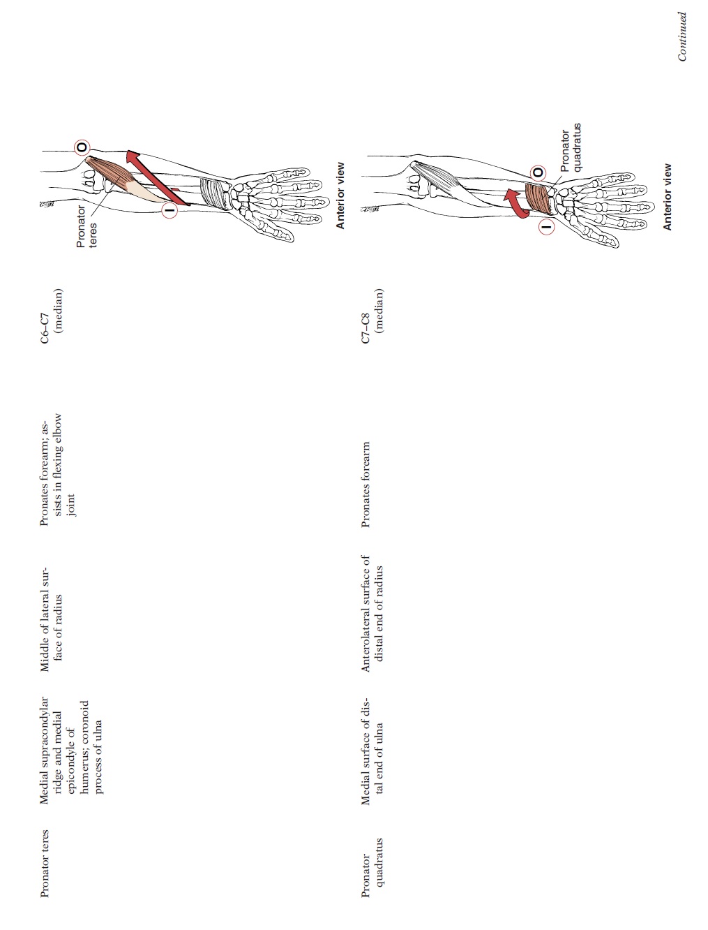
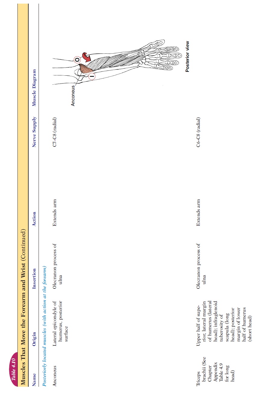
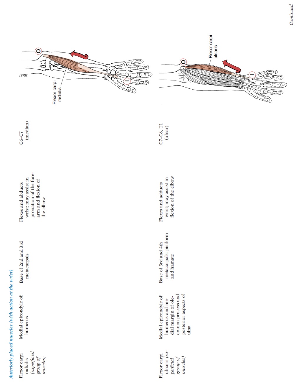
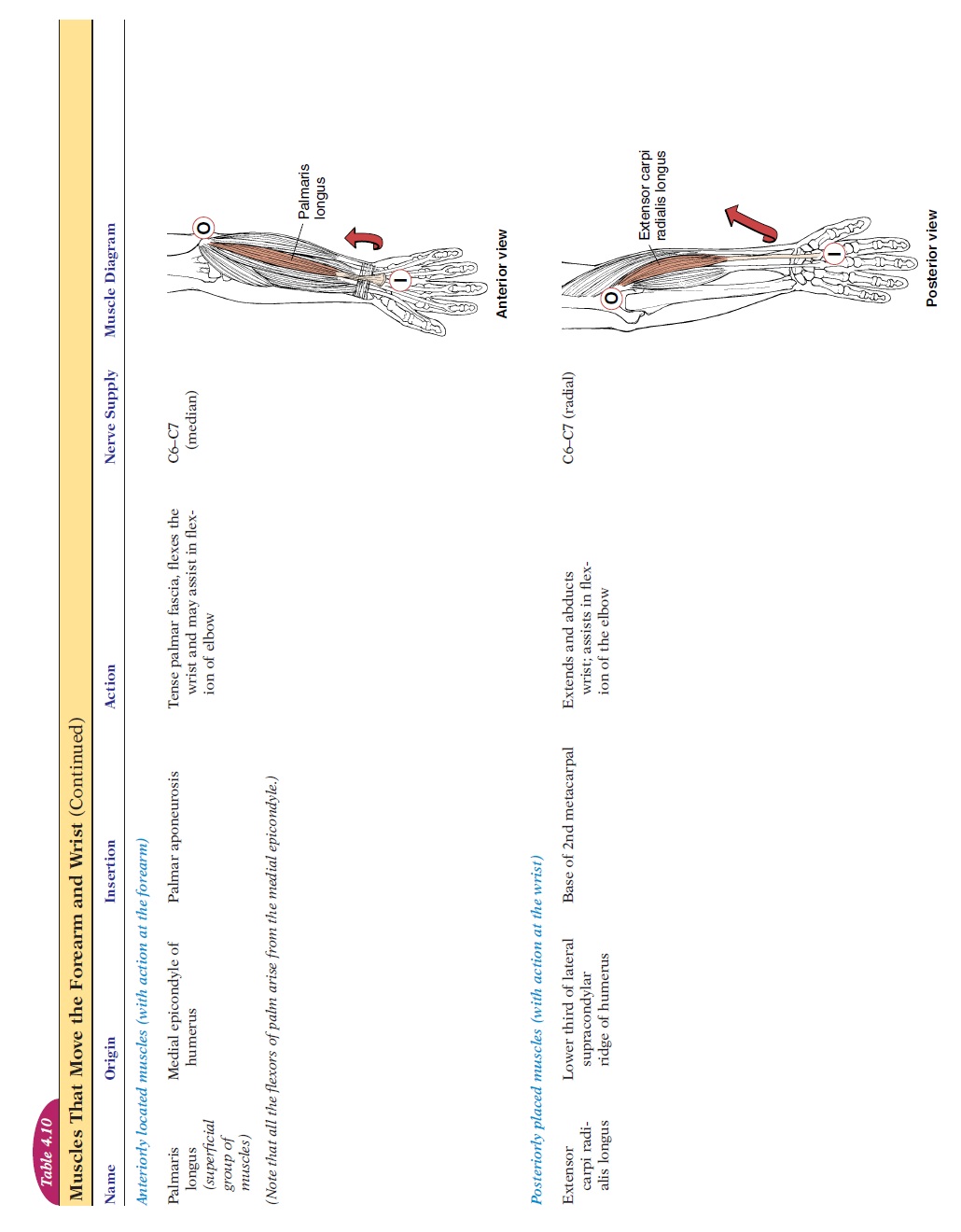
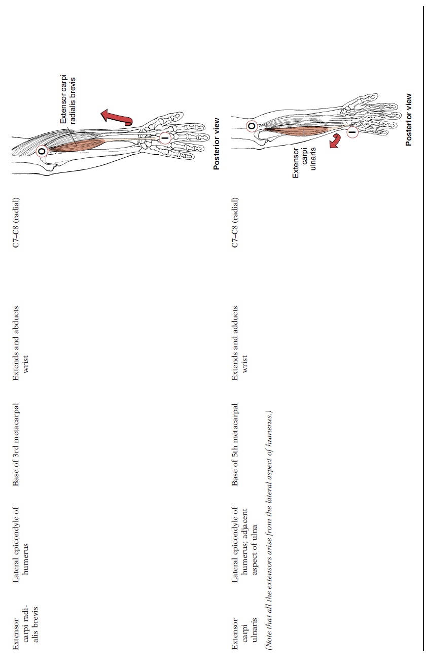
Muscles That Move the Forearm and Wrist
The muscles that move the forearm and wrist (see Figure 4.29, 4.10) gen-erally have their origins in the humerus (except for biceps brachii and the triceps brachii) and cross the elbow and/or wrist joint. At the elbow, the muscles produce flexion and extension of the forearm. In addition, some muscles, by rotating the radius over the lower end of the ulna, pronate (palm faces pos-teriorly) and supinate (palm faces anteriorly) the forearm.
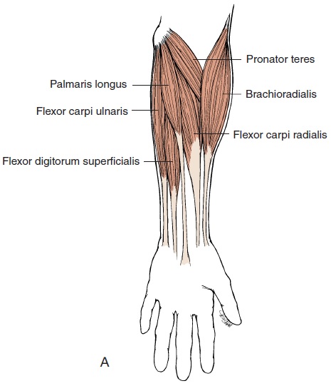
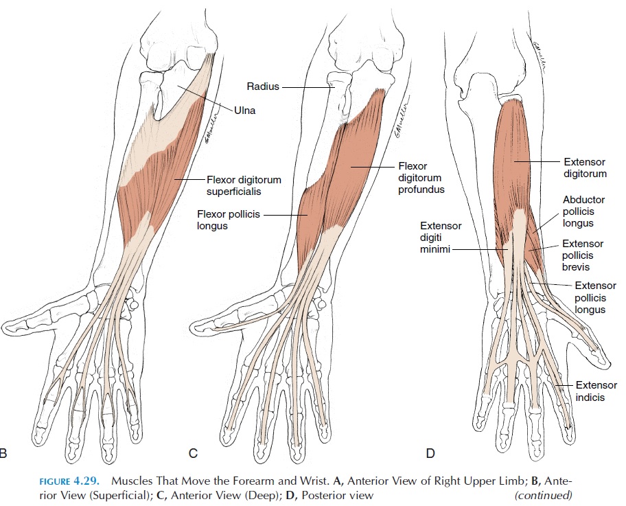
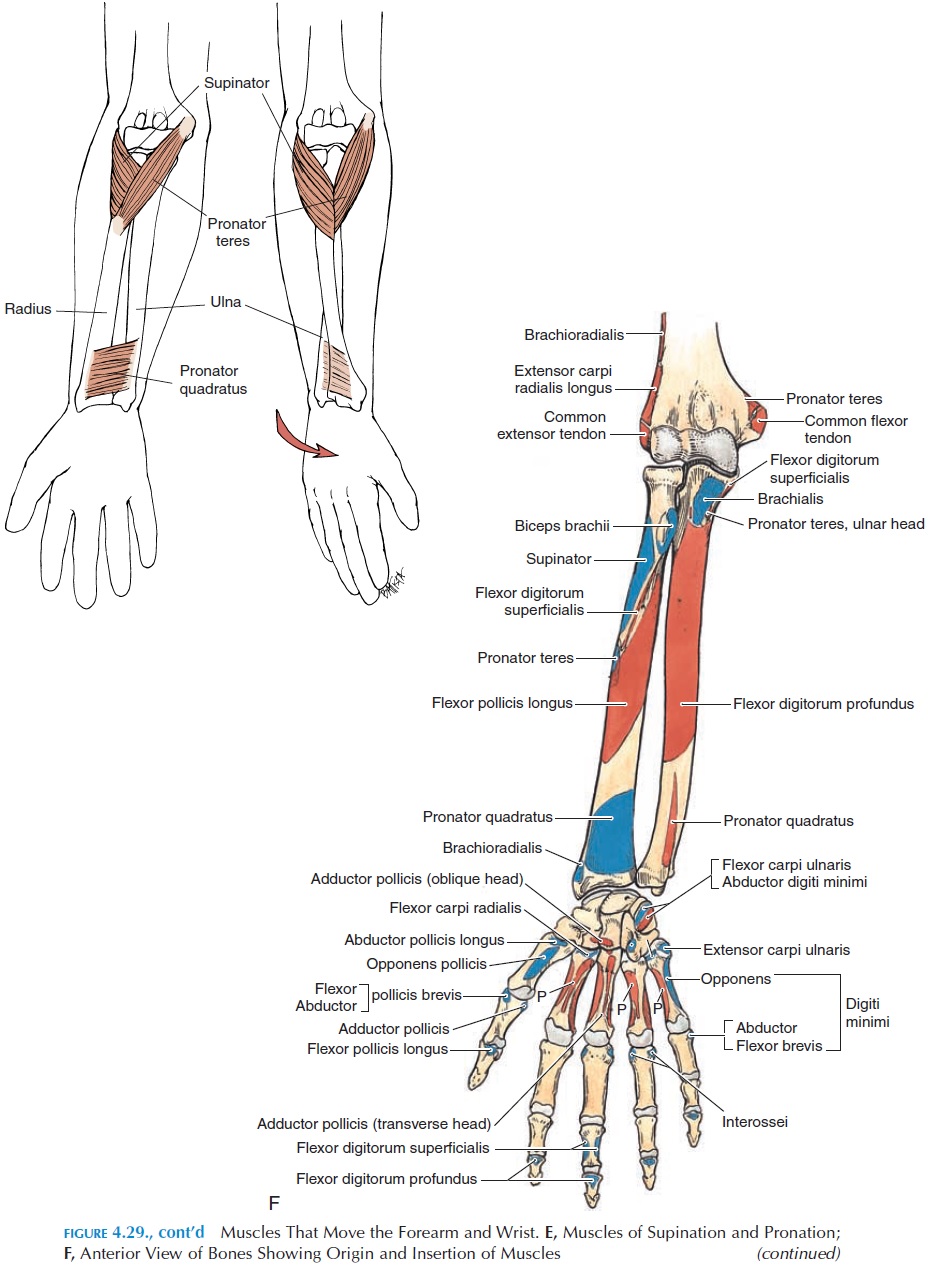
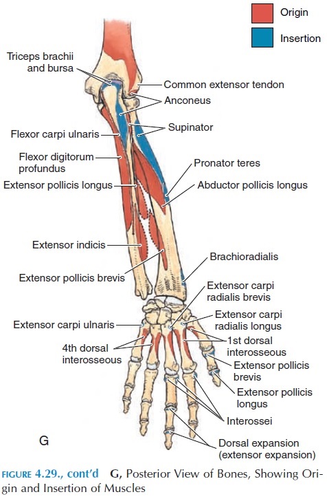
Flexion, extension, abduction, and adduction are movements that are brought about at the wrist. Notethat all the extensors arise on the lateral aspect of humerus.
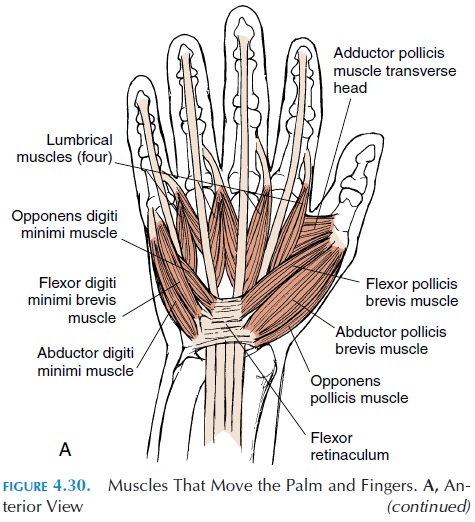
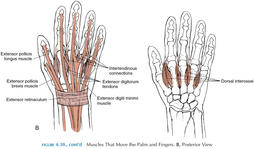
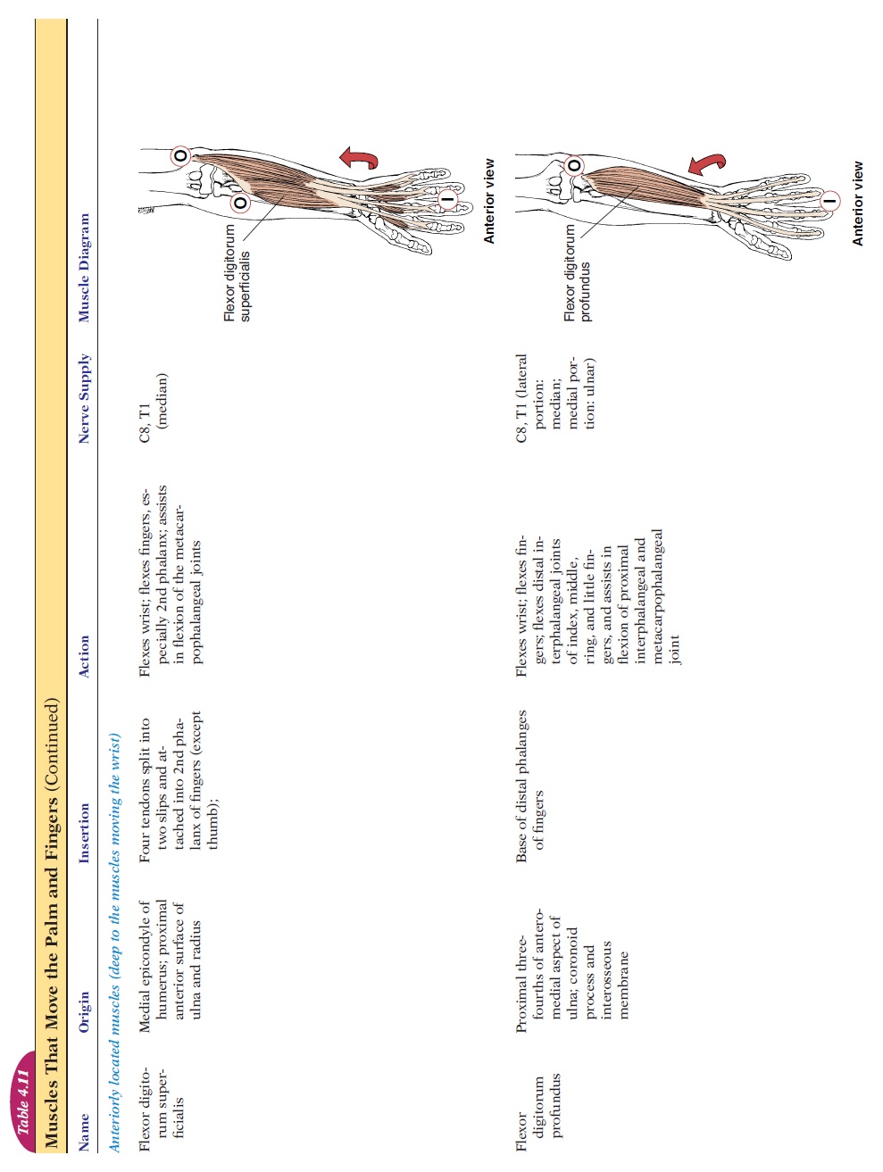
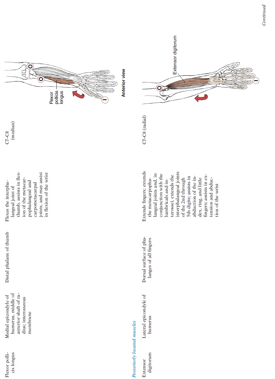
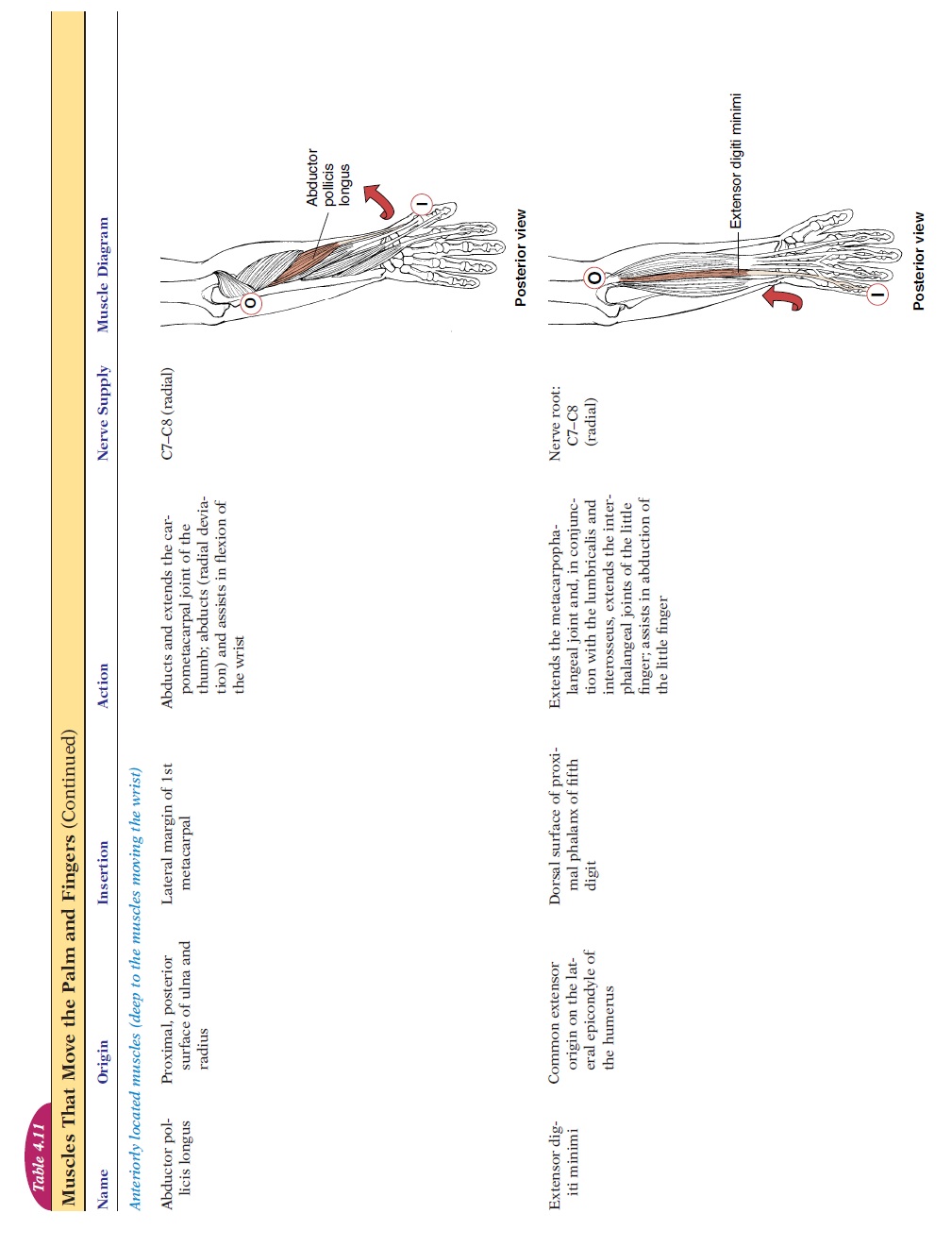
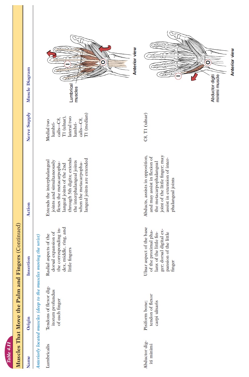
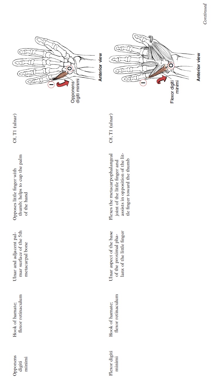
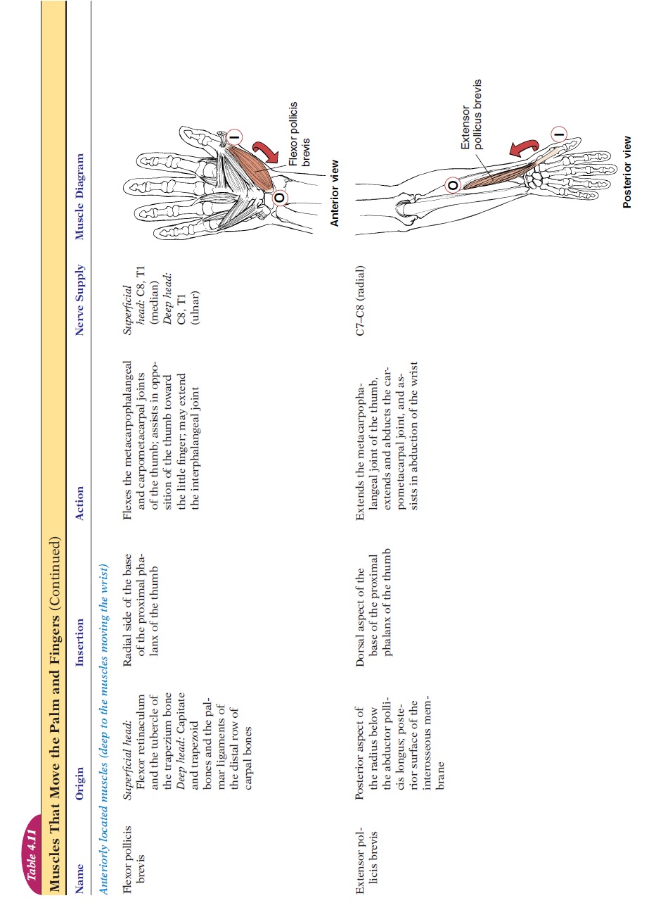
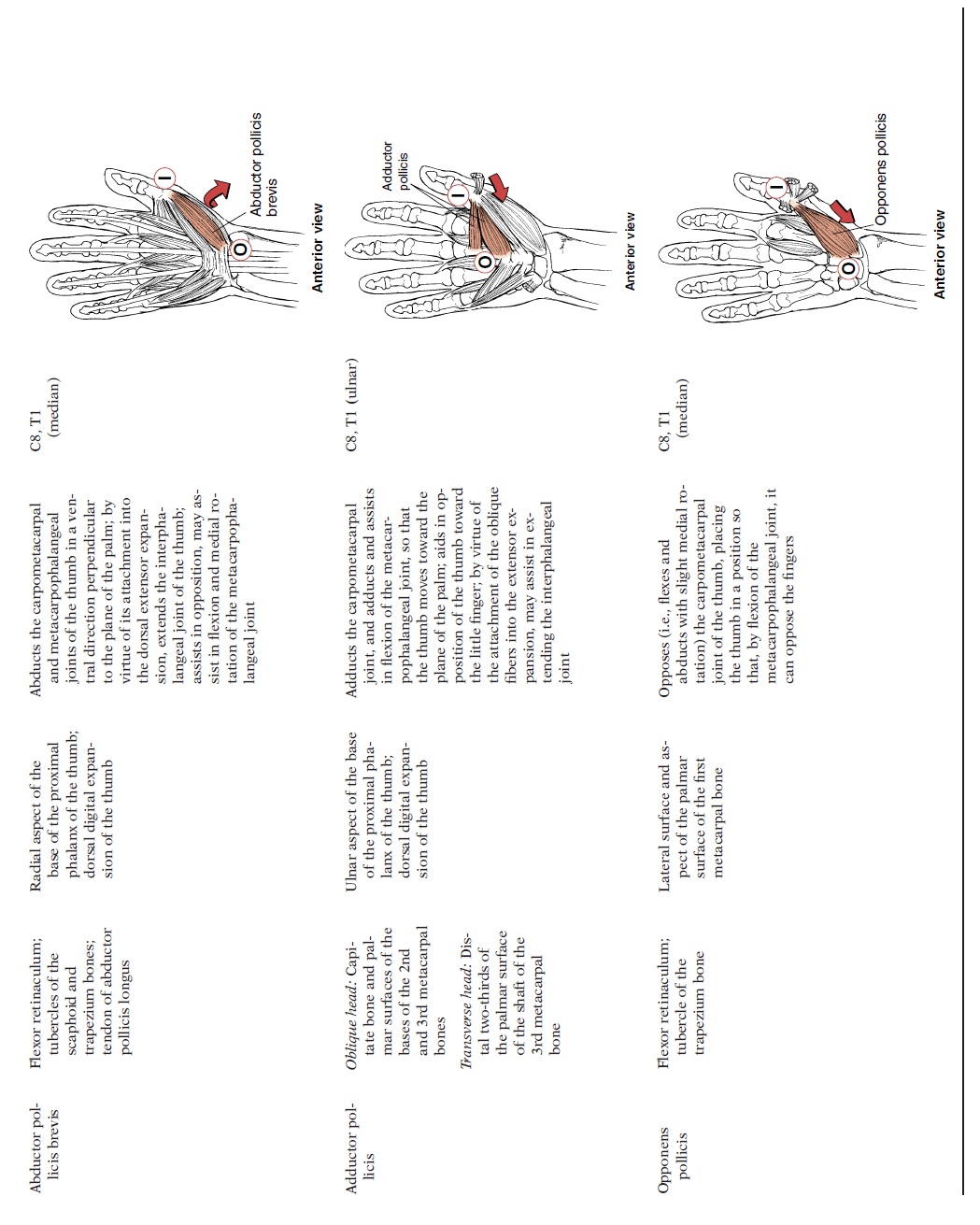
Muscles That Move the Palm and Fingers
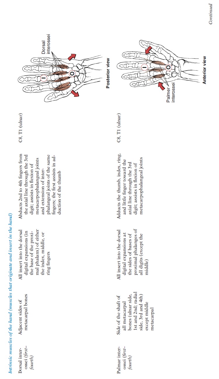
Many muscles involved in moving the palm and fin-gers (see Figure 4.30 and Table 4.11) are located in the forearm, with just the tendons extending onto the palm. This is an efficient way of increasing finger mobility and maintaining strength. Imagine how bulky the hand would be if all the mus-cles controlling the fingers arose in the palm! These muscles, the extrinsic muscles, primarily provide strength and are responsible for crude control of the hand. For finer control, small muscles arising from the carpals and metacarpals are known as the intrin-sic muscles of the hand.
Carpal Tunnel
The tendons of the muscles rising from the forearm are held in place against the carpals by connective tis sue sheets called retinaculum. The flexors are held in place by the flexor retinaculum and the extensors by the extensor retinaculum. The flexor retinacu-lum is a stamp-sized sheet of connective tissue run-ning anteriorly across the carpals. It forms a tunnel— carpal tunnel—through which nine flexor tendons and the median nerve pass (see Figure 4.31 and Fig-ure 3.43). The carpal tunnel is a narrow, rigid pas-
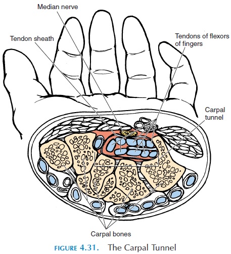
sage formed by the carpal bones of the wrist and the tough, inelastic transverse carpal ligament, or flexor retinaculum. As the tendons pass through the tunnel, they are surrounded by connective tissue sheaths (tendon sheaths or synovial sheaths) that are filled with synovial fluid. These sheaths reduce friction between the tendons as they move, lying close together in the wrist area. The median nerve in the carpal tunnel carries impulses to the muscles of the thumb and sensations from the skin over the thumb and the palmar surface of the lateral three and a half fingers.
The intrinsic muscles of the hand adduct, abduct, flex, and extend the fingers. Muscles are also present that help oppose the thumb and little finger. The bel-lies of the muscles that specifically move the thumb form a bulge on the lateral side of the palm called the thenar eminence. Those moving the little finger forma smaller bulge called the hypothenar eminence.
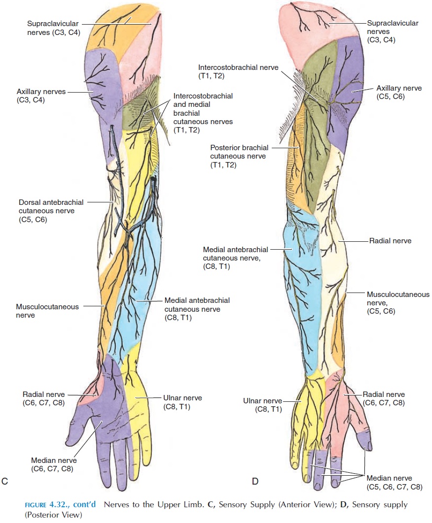
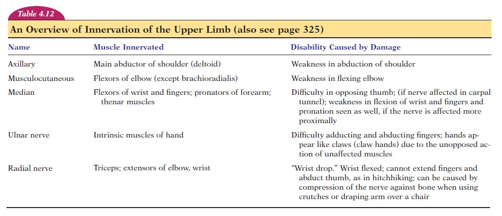
An Overview of Innervationof the Upper Limb
The muscles of the upper limb (see Figure 4.32 and Table 4.12) are innervated by nerves that arise from the cervical and upper thoracic segments of the spinal cord: C5–C8 and T1 (with con-tributions from C4 and T2). The nerve fibers (axons from these segments) form a network in the neck called the brachial plexus. From this network, after dividing and subdividing, five large nerves (nerve fiber bundles) are formed that go down the arm to innervate the muscles. The nerves are the axillary, musculocutaneous, median, ulnar, and radial.
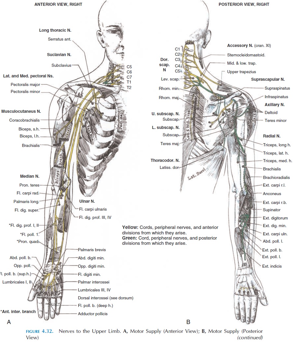
A general description of the nerve supply of these muscles is given in Table 4.12.

Related Topics