Chapter: The Massage Connection ANATOMY AND PHYSIOLOGY : Muscular System
Muscles Of The Lower Limb - Origin and Insertion of Muscles
MUSCLES OF THE LOWER LIMB
The muscles of the lower limb will be addressed in three functional groups: (1) muscles that move the thigh; (2) muscles that move the leg; and (3) muscles that move the foot and toes.
Muscles That Move the Thigh
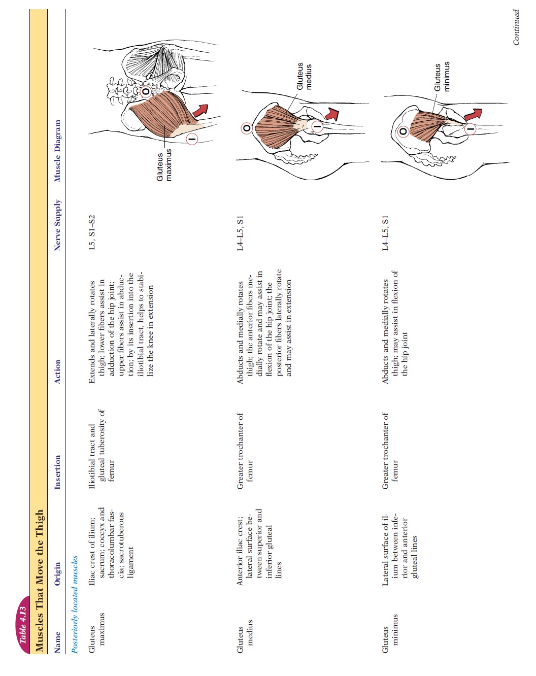
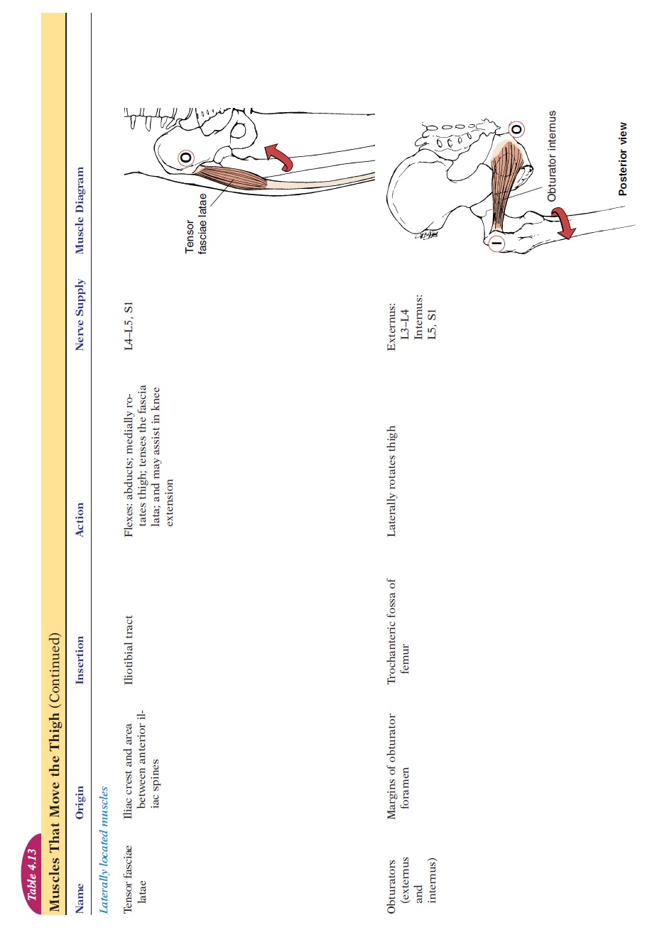
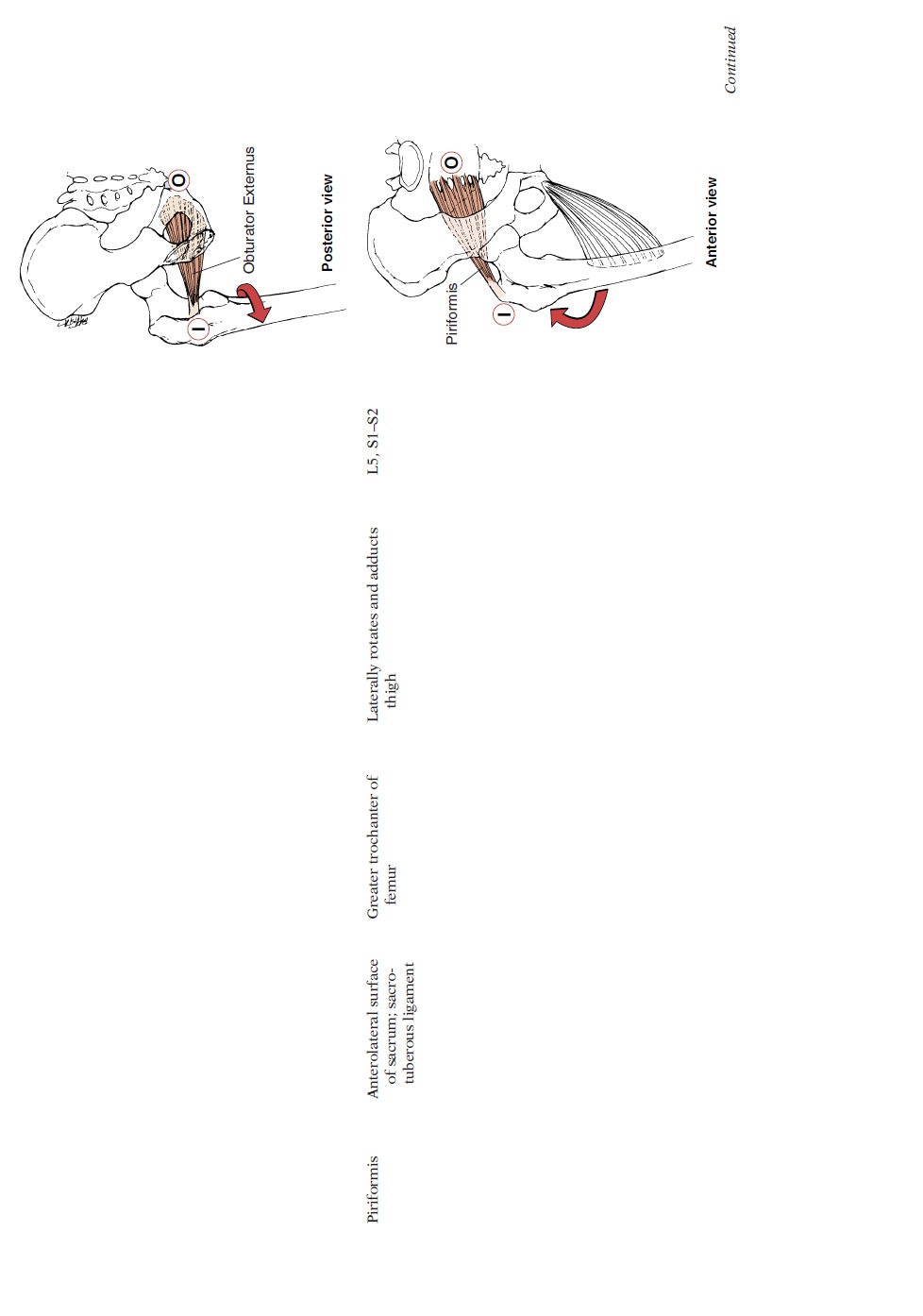
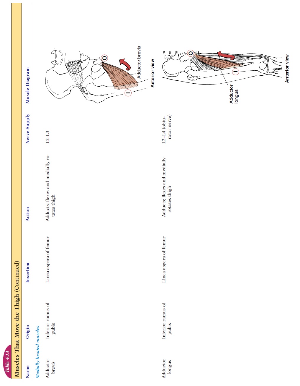
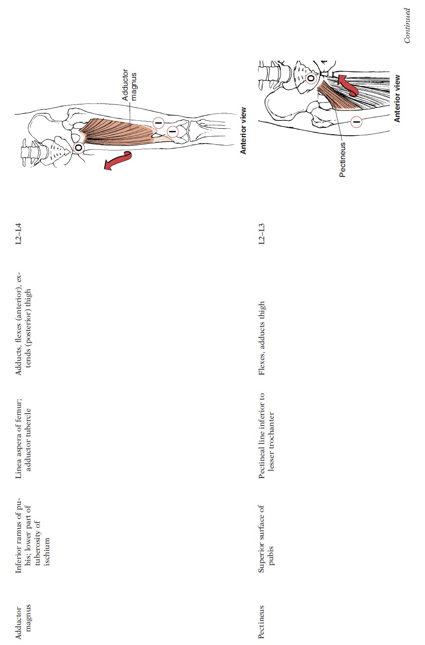
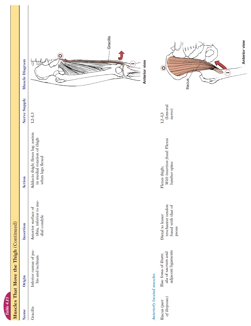
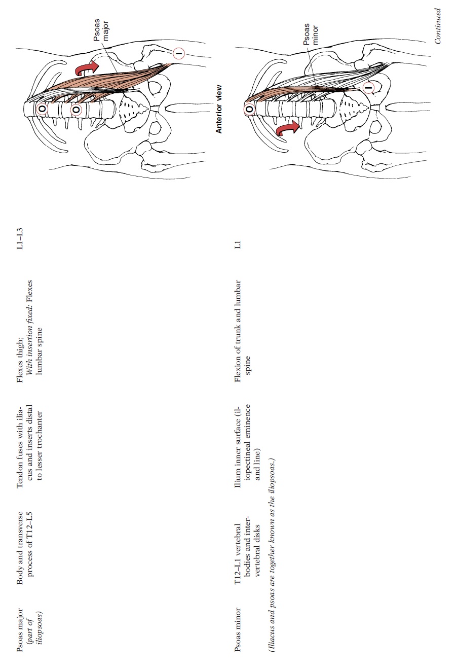
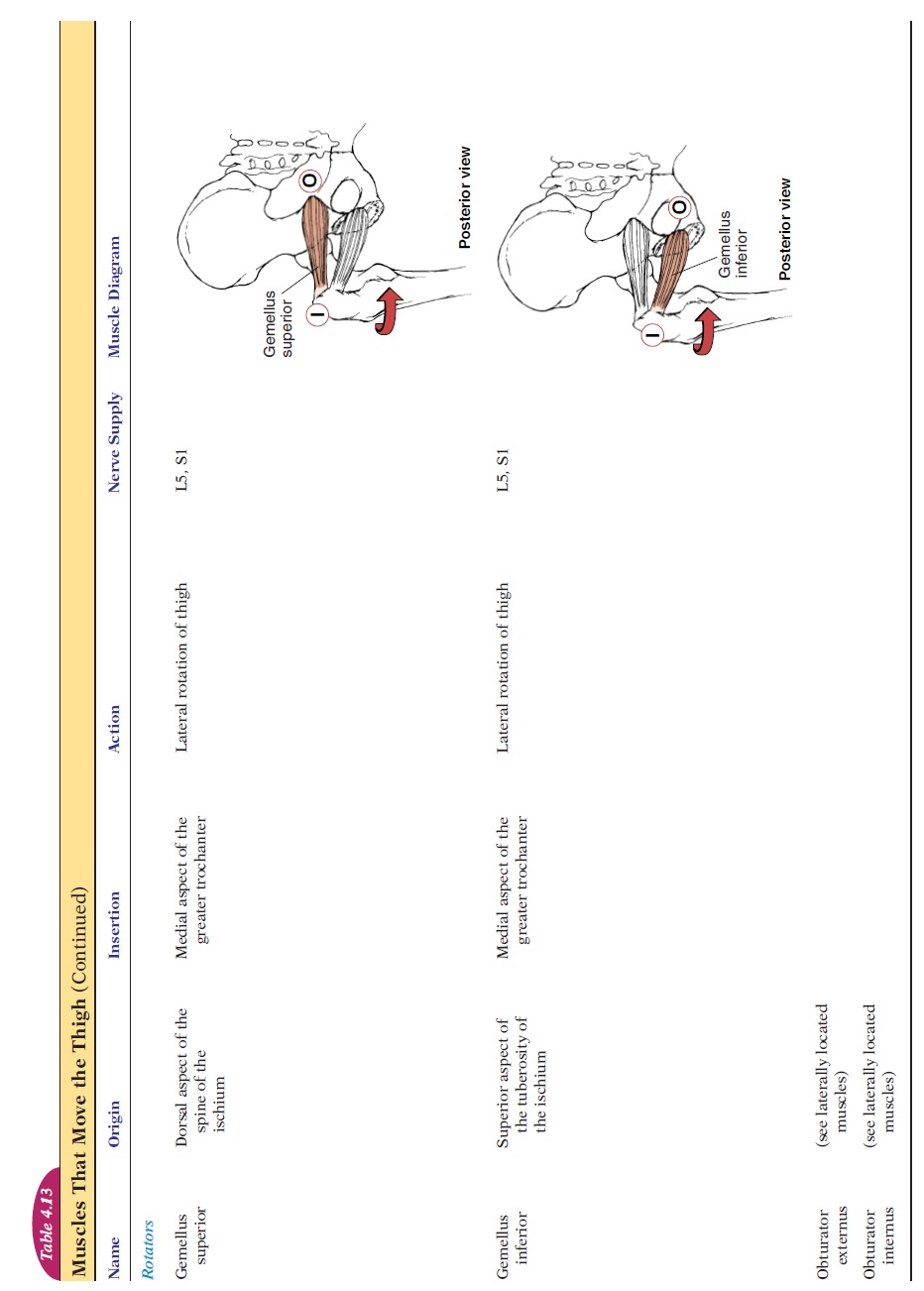
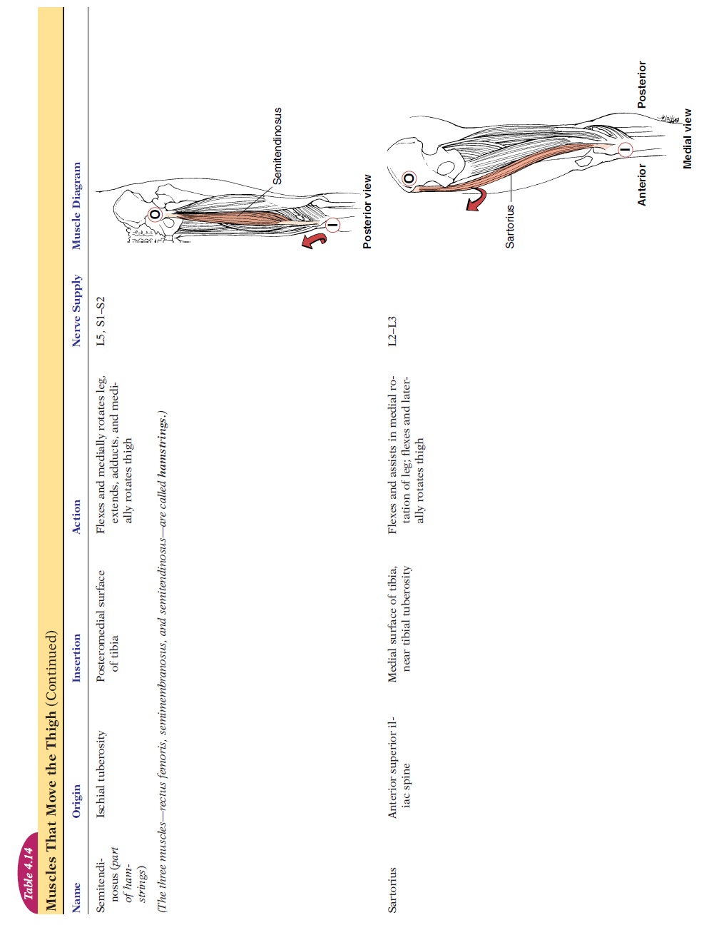
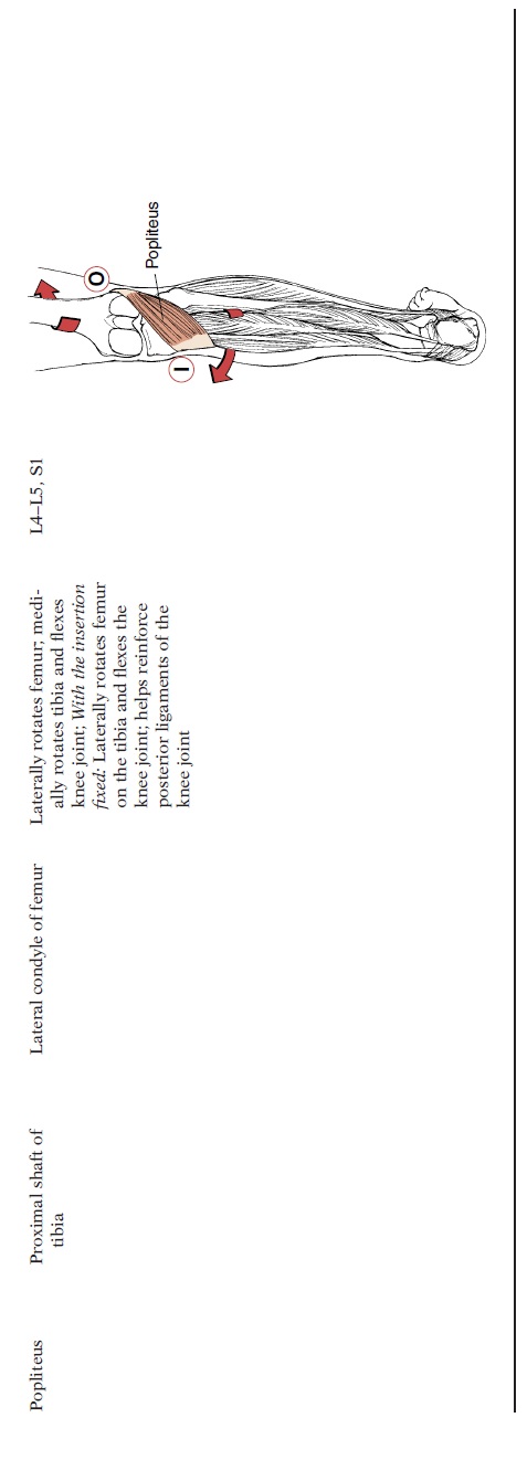
The muscles that move the thigh arise from the pelvis, except for the psoas that arises from the lower thoracic and lumbar vertebrae (see Figure 4.33 and Table 4.13). Those arising posteri-orly help extend the leg, and those arising anteriorly help flex the leg. Medially placed muscles (inner thigh) help adduction and those inserted laterally on the femur, help abduction. Muscles that are inserted medially and in the anterior aspect of the femur help rotate the leg medially. Those inserted into the lateral or posterior aspect of the femur produce lateral rotation. By knowing the origin and insertion of the muscles, the primary and secondary actions can be identified.
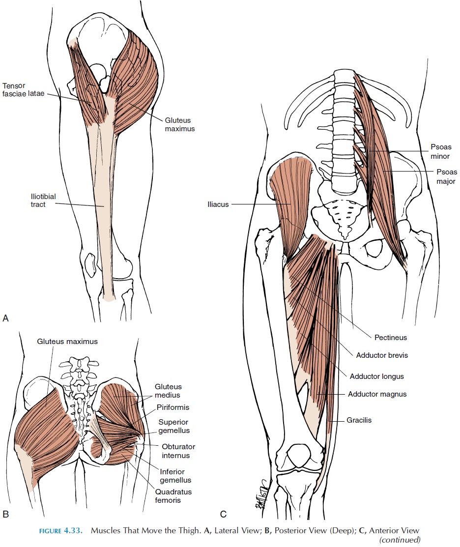
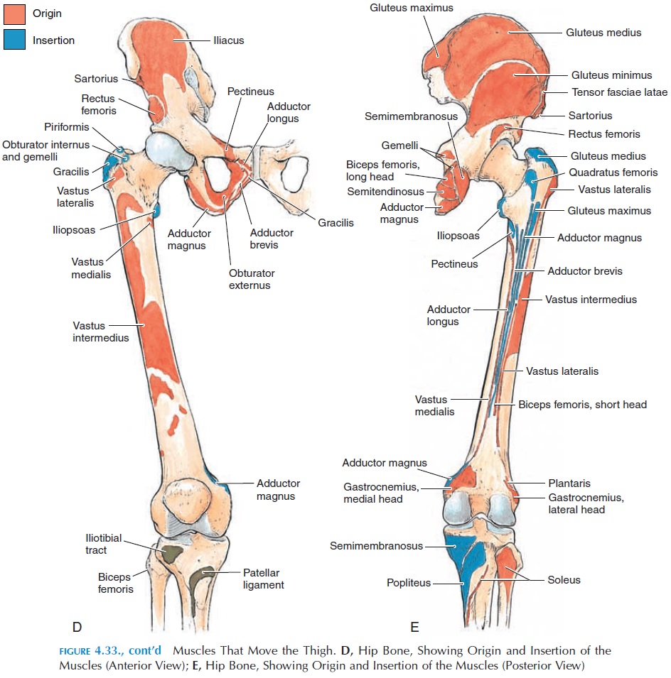
The gluteus maximus is the largest muscle located posteriorly. It inserts into a thick connective tissue sheet, the iliotibial tract. This tract is responsible for the indentation produced in the lateral part of the thigh when standing. The tract inserts into the upper end of tibia and helps brace the knee laterally. Table 4.13 shows the origin, insertion, and action of the muscles that move the thigh.
Muscles That Move the Leg
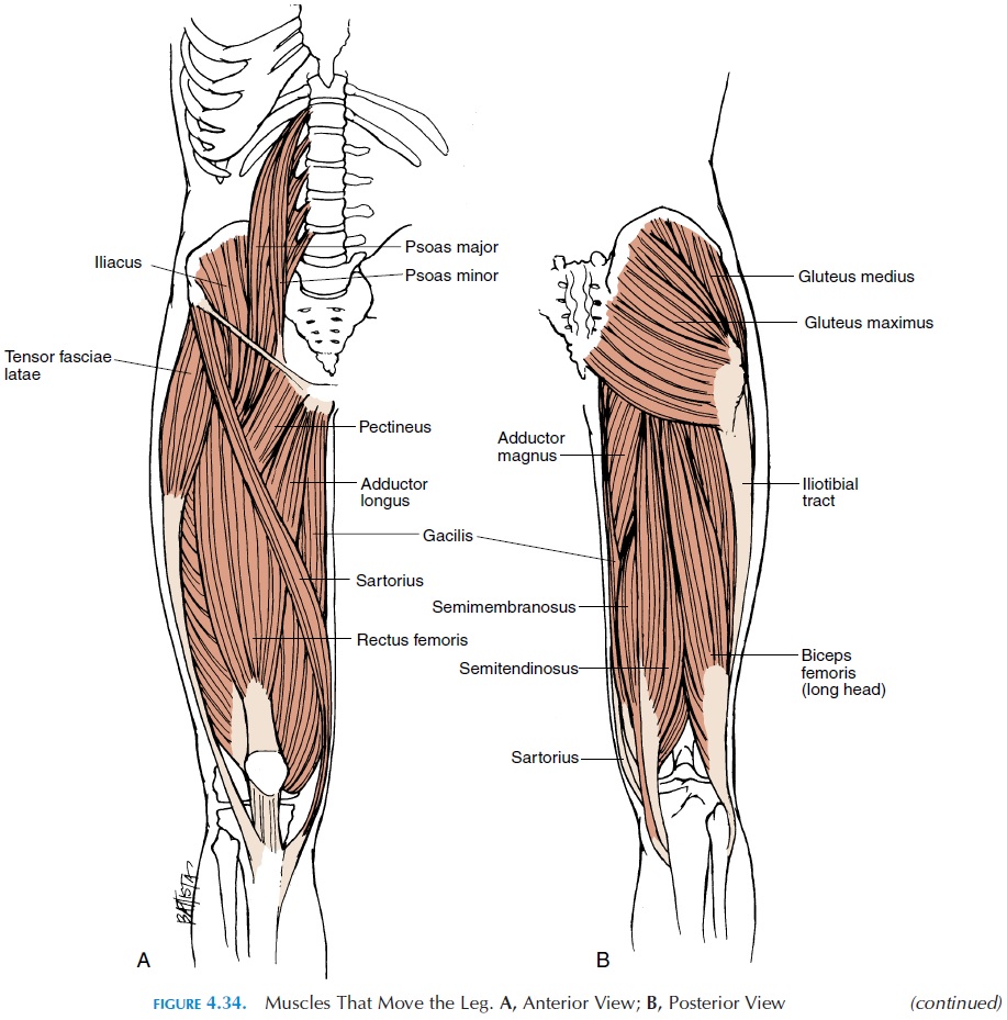
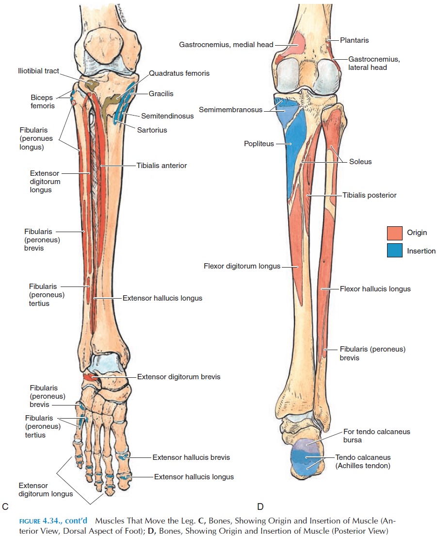
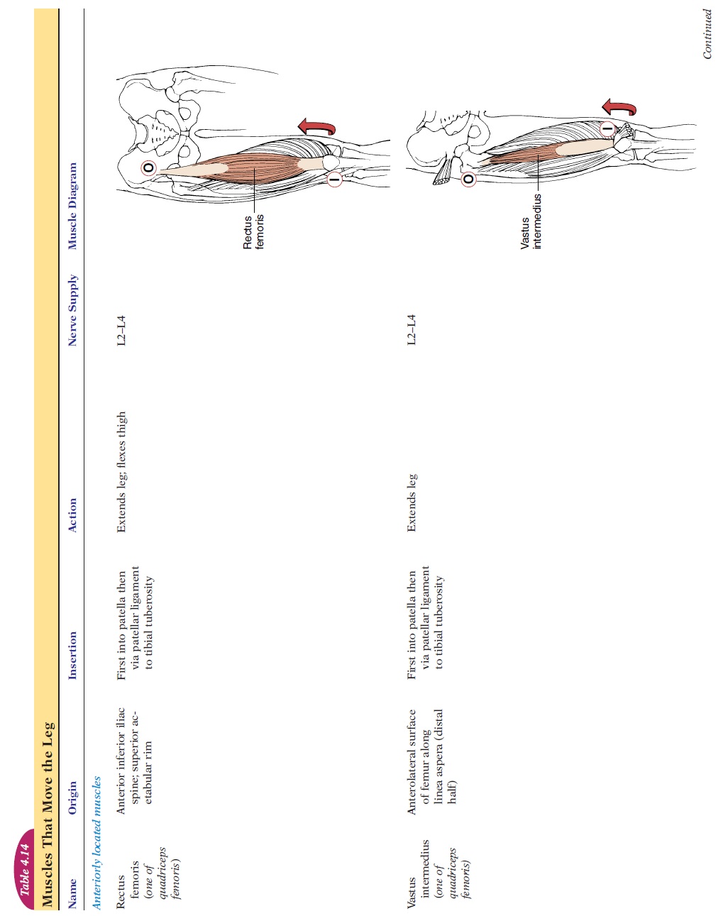
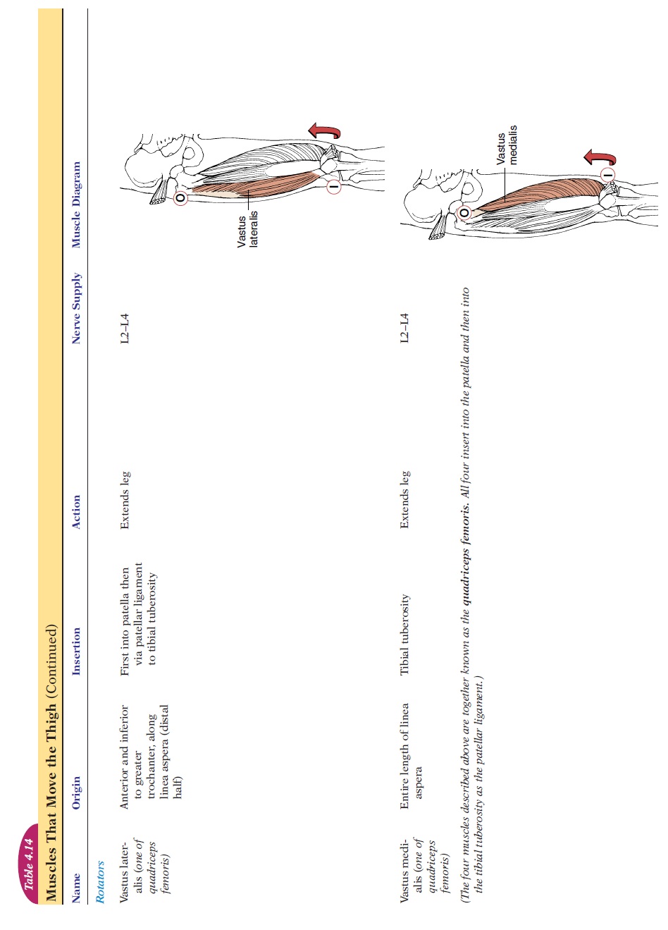
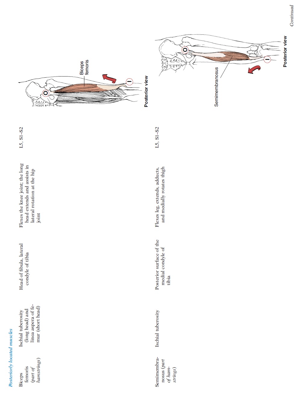
The arrangement of muscles in the lower limb is sim-ilar to that of the upper limb (see Figure 4.34 and Table 4.14). It must be remem-bered that flexion at the knee results in moving the lower leg posteriorly, unlike the upper limb where flexion of the elbow results in anterior movement of the forearm. The extensor group of muscles is located in the anterior and lateral aspect of the thigh; the flex-ors are located posteriorly and medially.
Muscles That Move the Foot and Toes
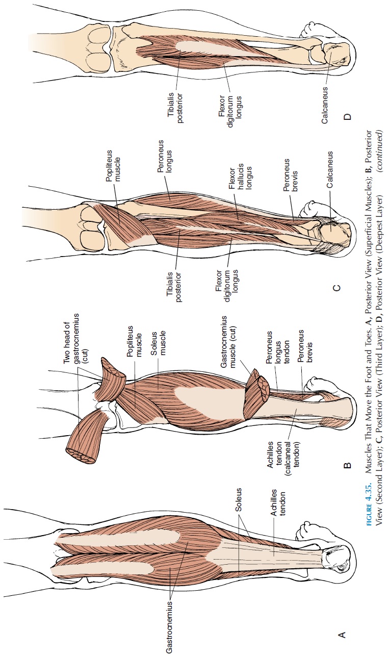
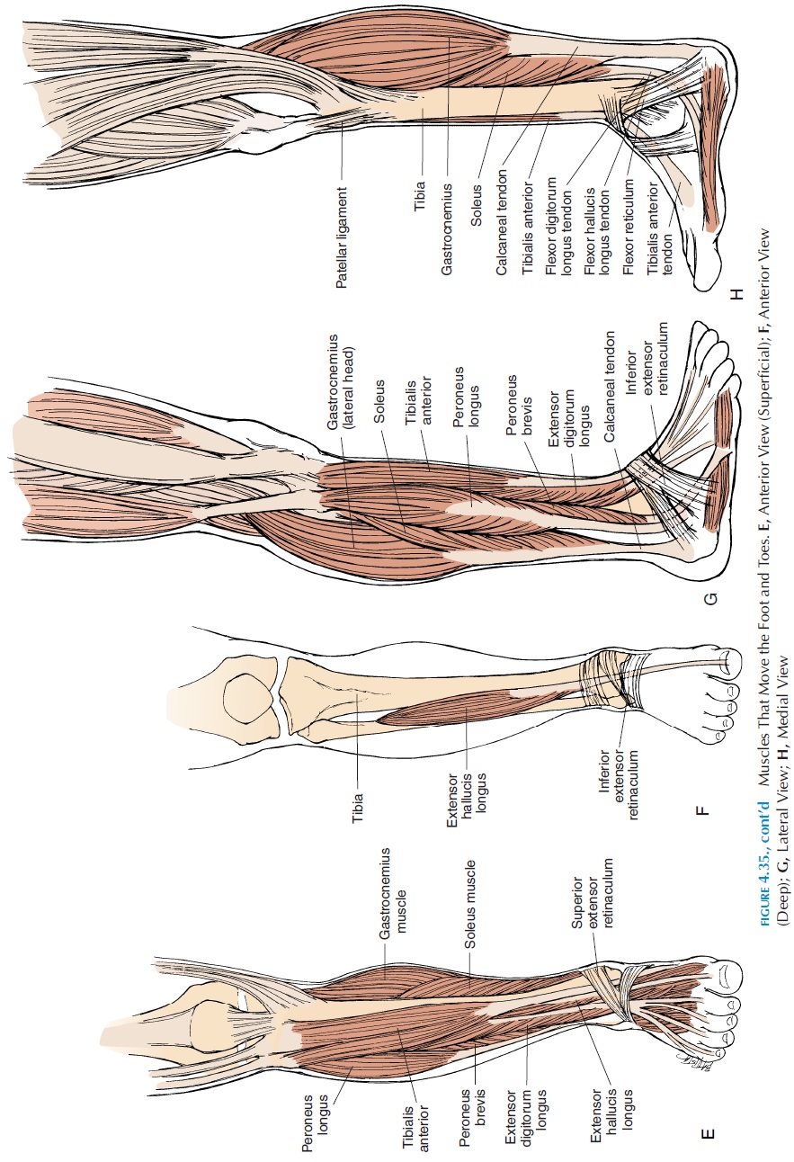
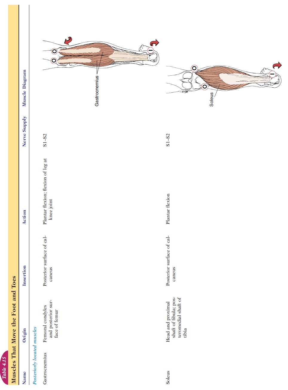
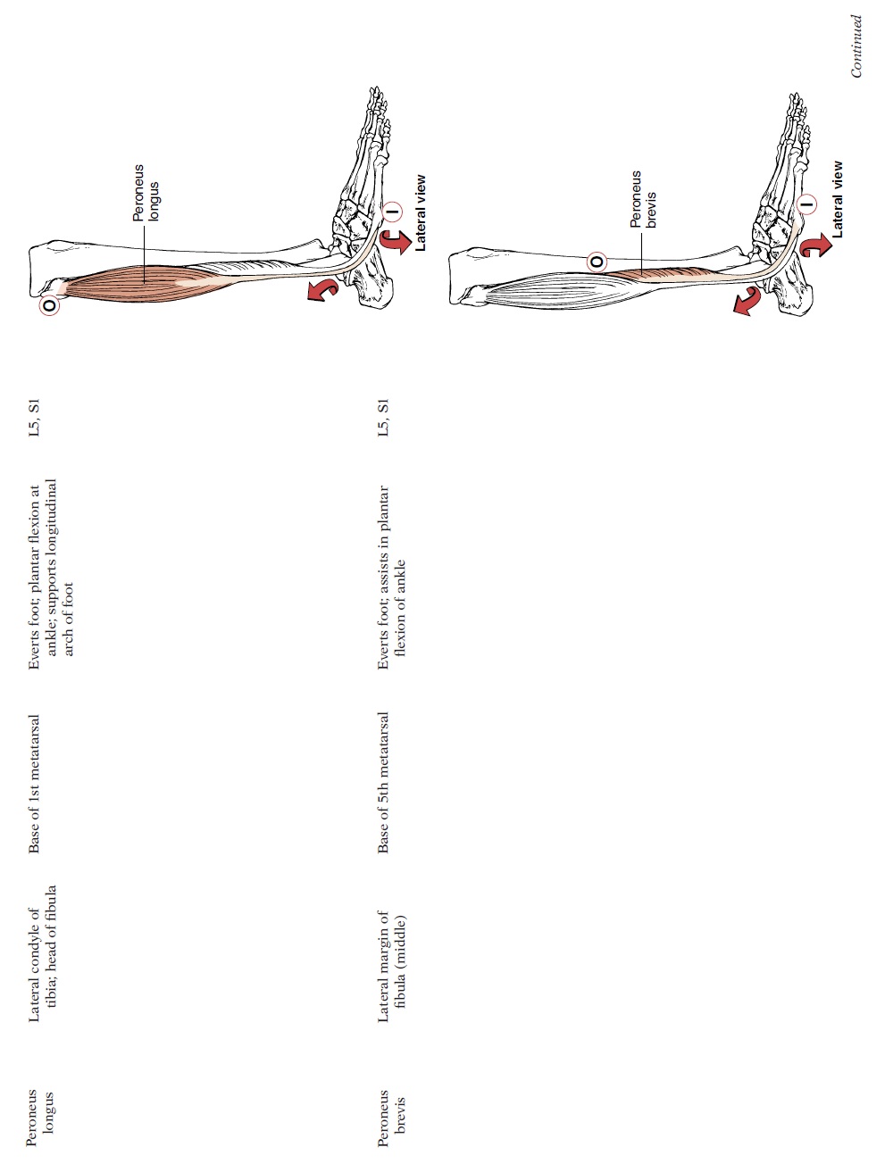
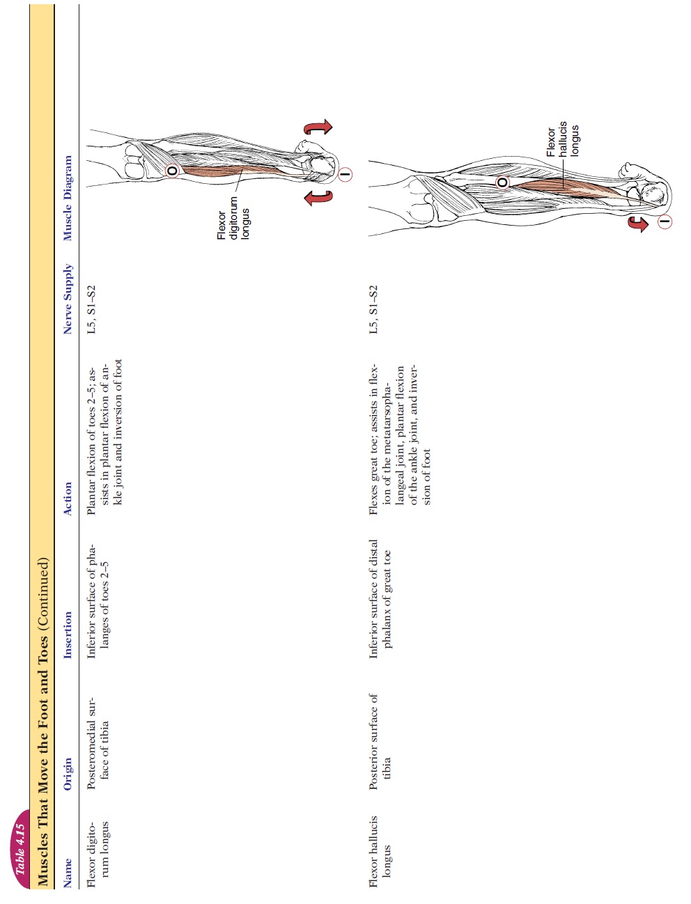
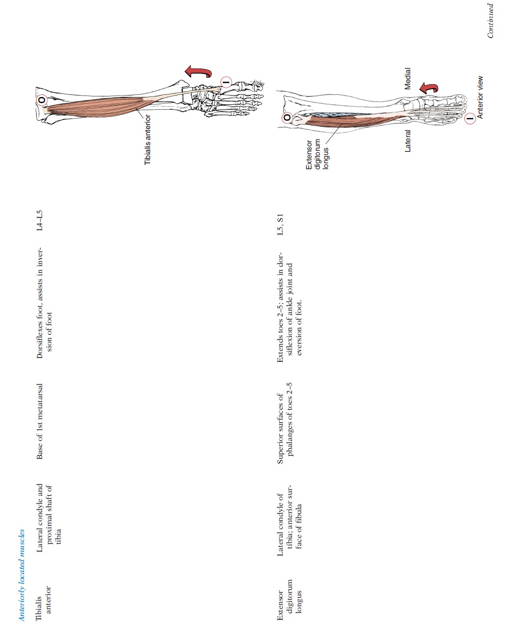
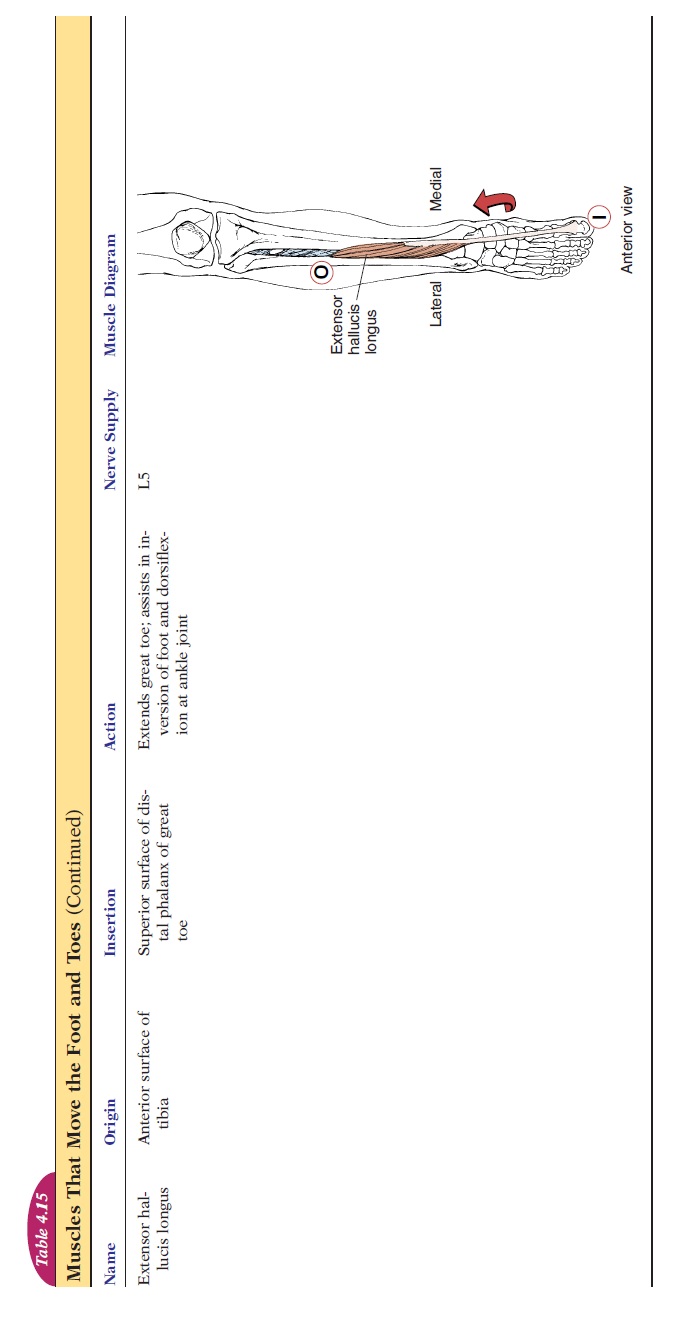
As in the forearm, extrinsic muscles of the foot are lo-cated in the anterior and posterior aspect of the tibia (see Figure 4.35 and Table 4.15). Those located posteriorly help with plantar flexion, and the muscles located anteriorly help extend or dorsiflex the foot. The large muscles located in the posterior aspect of the calf form a strong, thick ten-don called the Achilles tendon, or tendo calcaneus or calcaneal tendon. It is formed by the fusion of the soleus and gastrocnemius muscles.
Intrinsic Muscles of the Toes
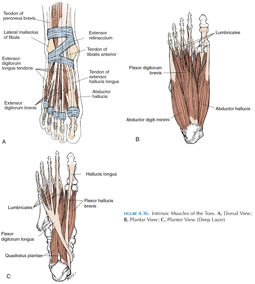
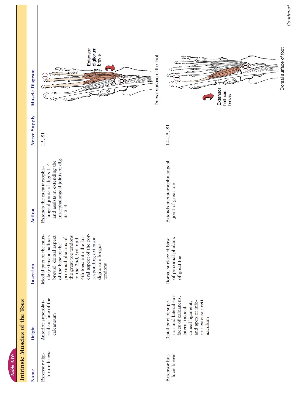
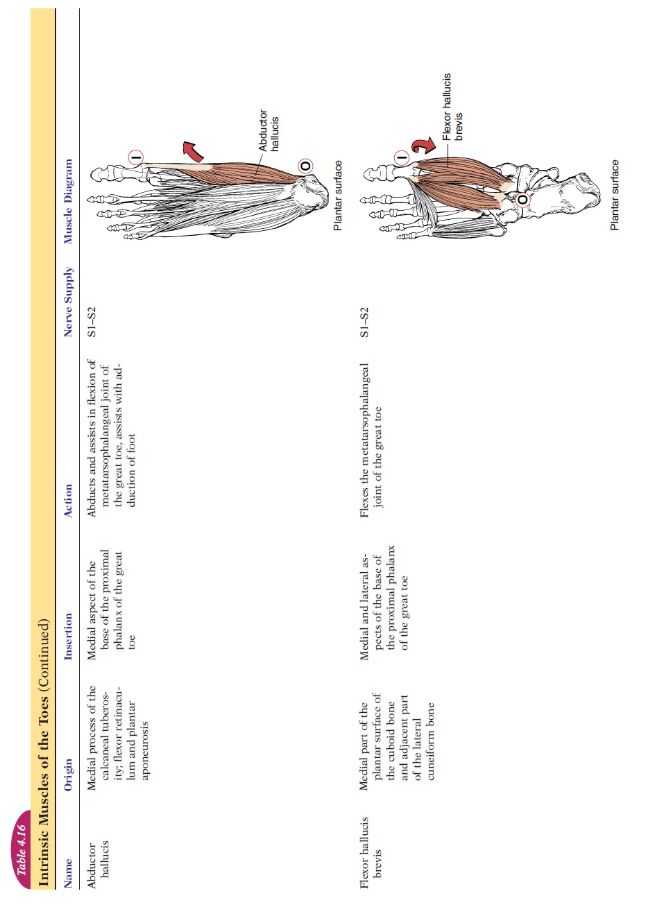
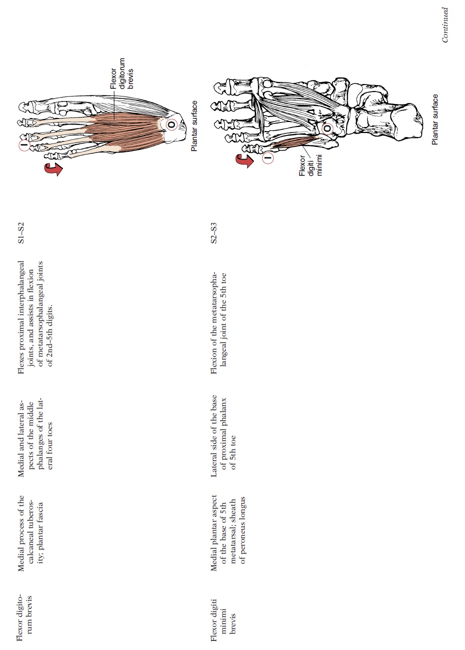
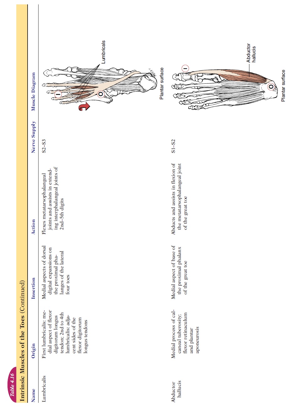
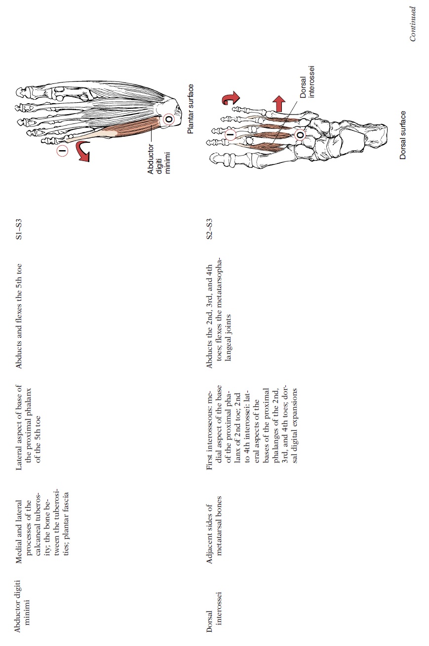
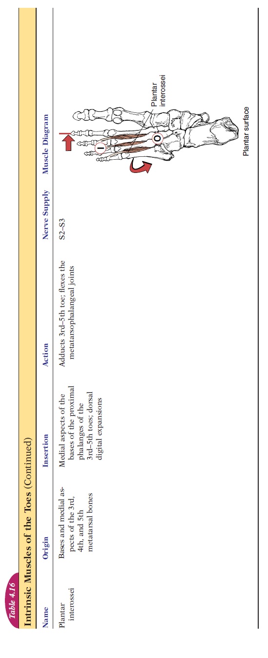
Like the intrinsic muscles of the hand, numerous mus-cles help adduct, abduct, flex, and extend the toes (see Figure 4.36 and Table 4.16). They arise from the tarsals and insert into the phalanges.
An Overview of Lower Limb Innervation
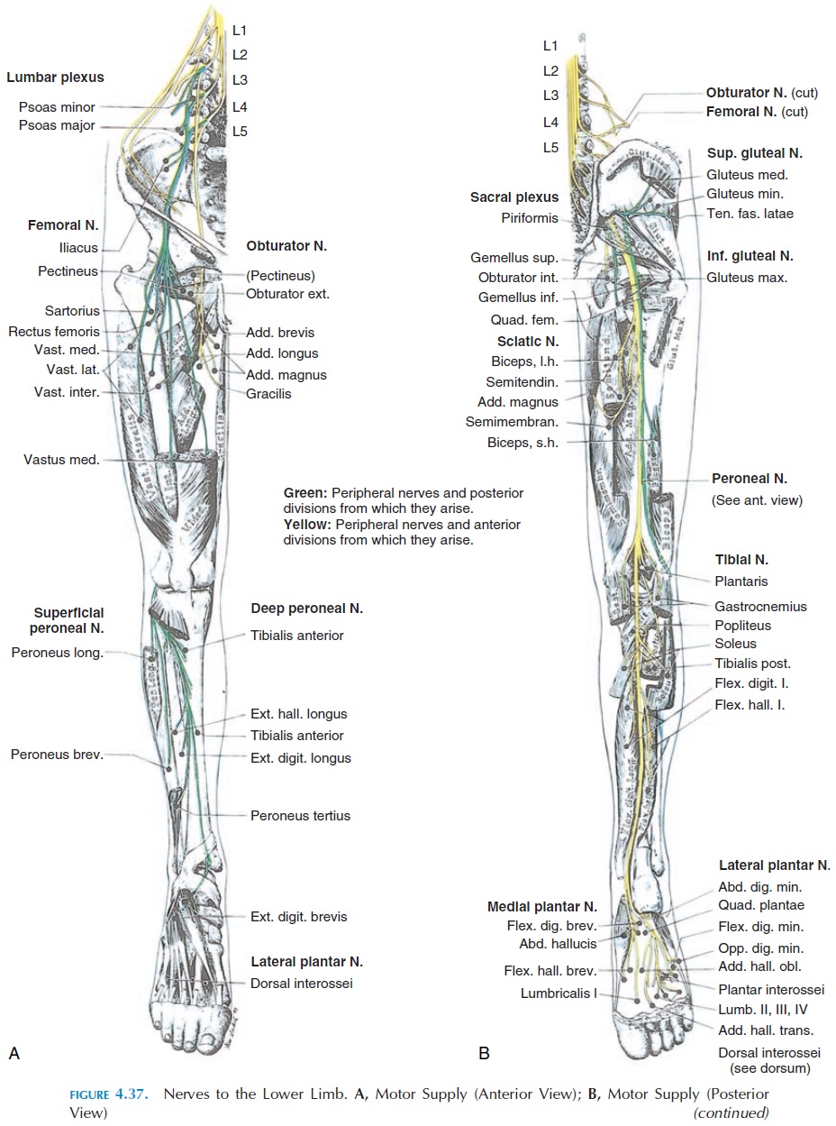
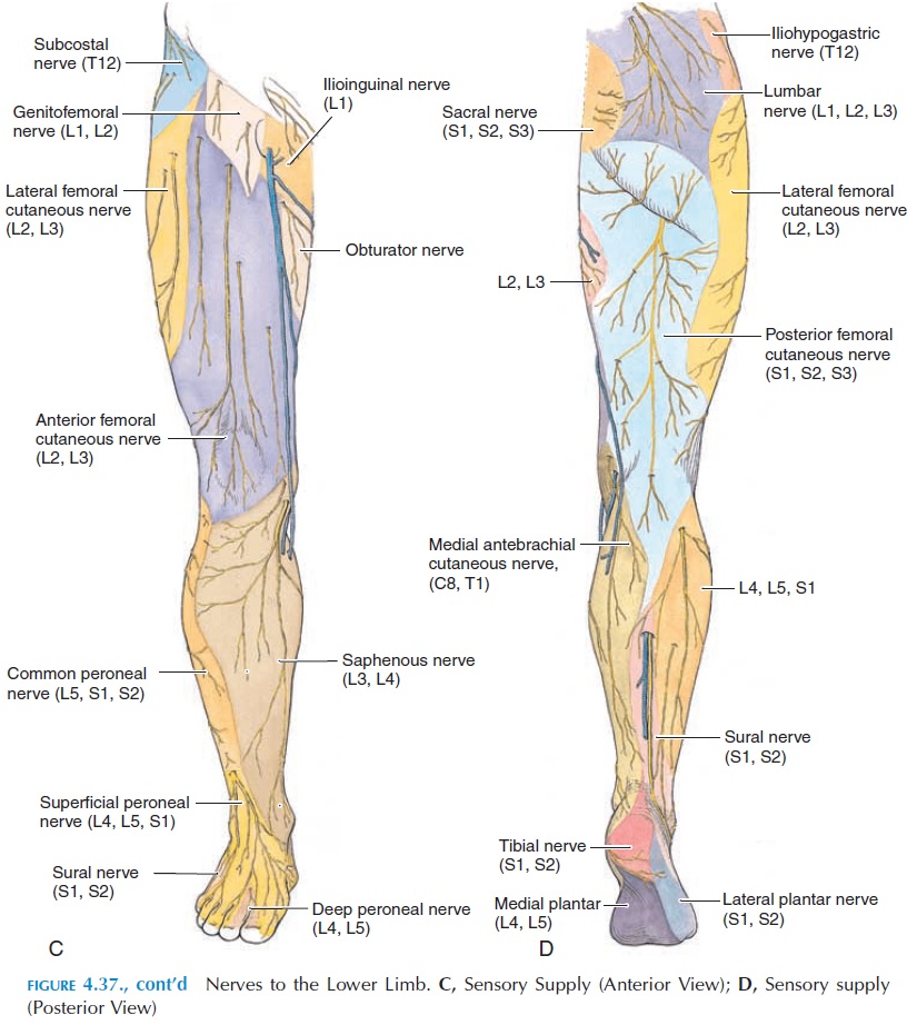
The muscles of the lower limb are innervated by nerves that arise from the lumbar (with contribution from T12, L1–L4) and sacral (L4–L5, S1–S3) segments of the spinal cord. These nerves initially form a net-work known as the lumbosacral plexus. Of the many nerves that arise from this plexus(see Figure 4.37), the sciatic nerve (L4–L5, S1–S3) is the largest (it is the largest nerve in the body). The sci-atic nerve, with its two main divisions—tibial and common peroneal nerves—supplies the skin of mostof the leg and foot, muscles of the posterior thigh, and all the muscles of the leg and foot.
Related Topics