Chapter: Ophthalmology: Glaucoma
Secondary Angle Closure Glaucoma

Secondary Angle Closure Glaucoma
Definition
In secondary angle closure glaucoma as in
primary angle closure glaucoma, the increase in intraocular pressure is due to
blockage of the trabecular mesh-work. However, the primary configuration of the
anterior chamber is not the decisive factor.
The most important causes:
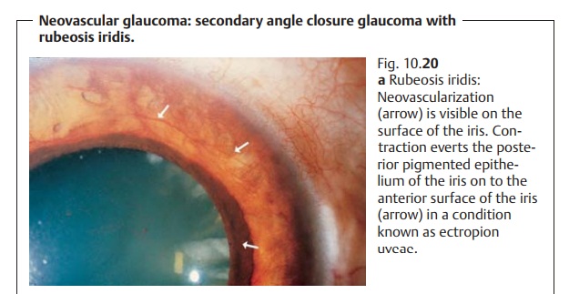
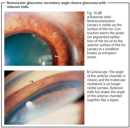
Rubeosis iridis.Neovascularization draws theangle of the anterior chamber
together like a zipper (neovascular glaucoma).
Ischemic retinal disorders such as diabetic retinopathy and retinal vein occlu-sion can lead to
rubeosis iridis with progressive closure of the angle of theanterior chamber.
Other forms of retinopathy or intraocular tumors can also cause rubeosis
iridis. The prognosis for eyes with neovascular glaucoma is poor (see Fig. 10.20a and b).

Trauma.Post-traumatic presence of blood or exudate in
the angle of the ante-rior chamber and prolonged contact between the iris and
trabecular mesh-work in a collapsed anterior chamber (following injury,
surgery, or insuffi-cient treatment of primary angle closure) can lead to anterior
synechiae and angle closure without rubeosis iridis.
Medical therapy of secondary glaucomas is
usually identical to the treatment of primary chronic open angle glaucoma.
Secondary glaucomas may be caused by many
different factors, and the angle may be open or closed. Therefore, treatment
will depend on the etiology of the glaucoma. The underlying disorder is best
treated first. Glaucomas with uveitis (such as iritis or iridocyclitis)
initially are treated conservatively with anti-inflammatory and antiglaucoma
agents. Surgery is indicated where con-servative treatment is not sufficient.
The prognosis for secondary glaucomas is
generally worse than for pri-mary glaucomas.
Related Topics