Chapter: Essentials of Anatomy and Physiology: Heart
Route of Blood Flow Through the Heart
Route Of Blood Flow Through The Heart
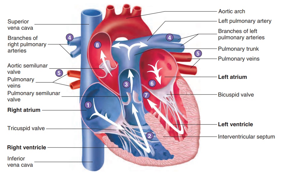
The route of blood flow through the heart is depicted in figure 12.10. Even though blood flow is described for the right and then the left side of the heart, it is important to understand that both atria contract at the same time, and both ventricles contract at the same time. This concept is most important when considering the electrical activity, pressure changes, and heart sounds.
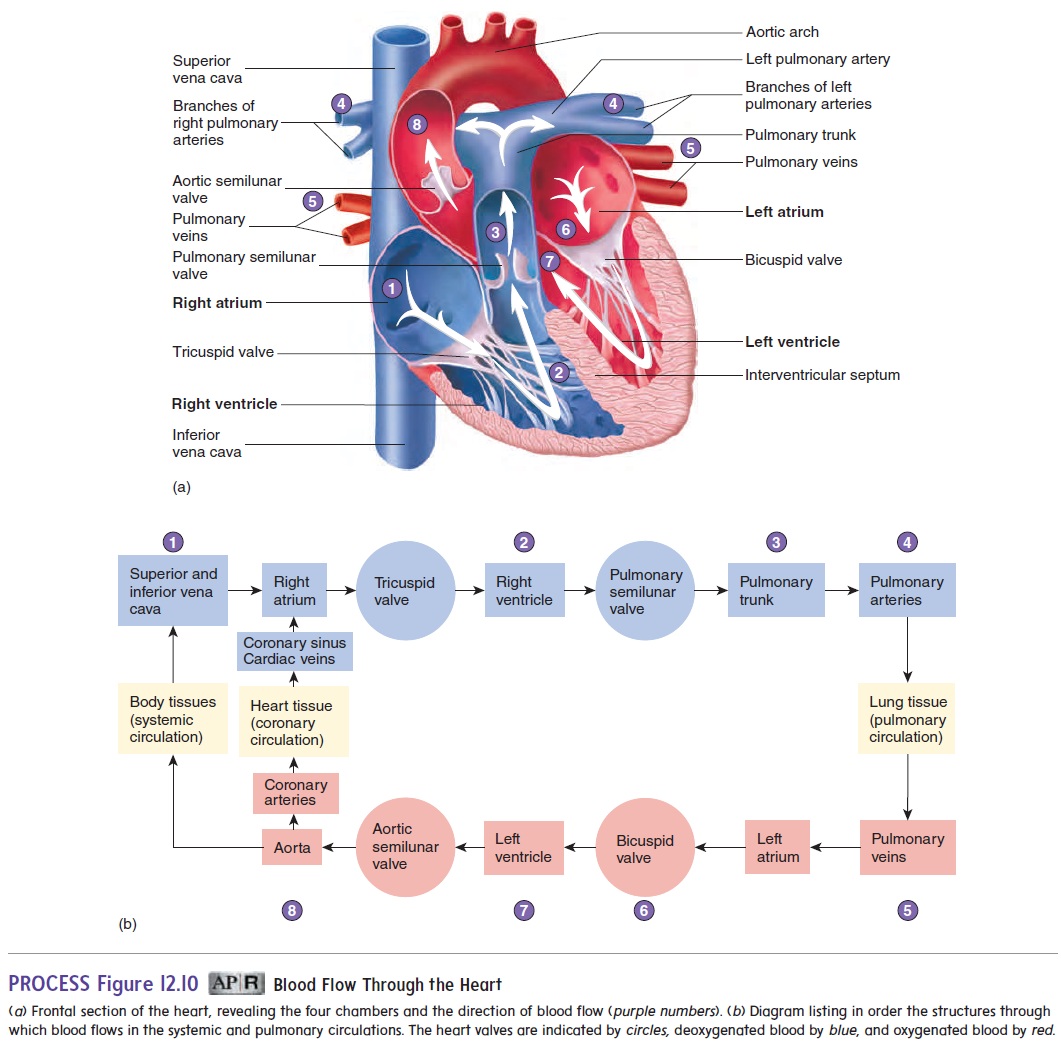
Blood enters the right atrium from the systemic circulation through the superior and inferior venae cavae, and from heart muscle through the coronary sinus. Most of the blood flowing into the right atrium flows into the right ventricle while the right ven-tricle relaxes following the previous contraction. Before the end of ventricular relaxation, the right atrium contracts, and enough blood is pushed from the right atrium into the right ventricle to complete right ventricular filling.
Following right atrial contraction, the right ventricle begins to contract. This contraction pushes blood against the tricuspid valve, forcing it closed. After pressure within the right ventricle increas-es, the pulmonary semilunar valve is forced open, and blood flows into the pulmonary trunk. As the right ventricle relaxes, its pressure falls rapidly, and pressure in the pulmonary trunk becomes greater than in the right ventricle. The backflow of blood forces the pul-monary semilunar valve to close.
The pulmonary trunk branches to form the right and left pulmonary arteries, which carry blood to the lungs, where CO2 is released and O2 is picked up. Blood returning from the lungs enters
Most of the blood flowing into the left atrium passes into the left ventricle while the left ventricle relaxes following the previous contraction. Before the end of ventricular relaxation, the left atrium contracts, and enough blood is pushed from the left atrium into the left ventricle to complete left ventricular filling.
Following left atrial contraction, the left ventricle begins to contract. This contraction pushes blood against the bicuspid valve, forcing it closed. After pressure within the left ventricle increases, the aortic semilunar valve is forced open, and blood flows into the aorta. Blood flowing through the aorta is distributed to all parts of thebody, except to those parts of the lungs supplied by the pulmonary blood vessels. As the left ventricle relaxes, its pressure falls rapidly, and pressure in the aorta becomes greater than in the left ventricle. The backflow of blood forces the aortic semilunar valve to close.
Related Topics