Chapter: Essentials of Anatomy and Physiology: Heart
Blood Supply to the Heart
Blood Supply to the Heart
Coronary Arteries
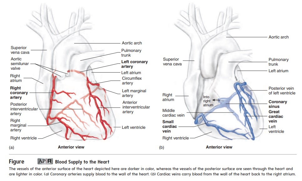
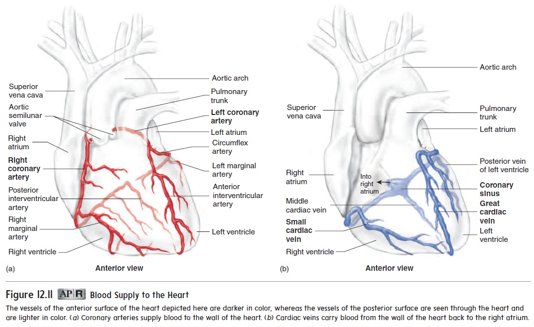
The cardiac muscle in the wall of the heart is thick and metabolically very active. Two coronary arteries supply blood to the wall of the heart (figure 12.11a). The coronary arteries originate from the base of the aorta, just above the aortic semilunar valves. The left coro-nary artery originates on the left side of the aorta. It has three majorbranches: The anterior interventricular artery lies in the anterior interventricular sulcus; the circumflex artery extends around the coronary sulcus on the left to the posterior surface of the heart; and the left marginal artery extends inferiorly along the lateral wall of the left ventricle from the circumflex artery. The branches of the left coronary artery supply much of the anterior wall of the heart and most of the left ventricle. The right coronary artery originates on the right side of the aorta. It extends around the coronary sulcus on the right to the posterior surface of the heart and gives rise to the posterior interventricular artery, which lies in the posterior inter-ventricular sulcus. The right marginal artery extends inferiorly along the lateral wall of the right ventricle. The right coronary artery and its branches supply most of the wall of the right ventricle.
In a resting person, blood flowing through the coronary arter-ies gives up approximately 70% of its O2. In comparison, blood flowing through arteries to skeletal muscle gives up only about 25% of its O2. The percentage of O2 the blood releases to skeletal muscle increases to 70% or more during exercise, but the percent-age of O2 the blood releases to cardiac muscle cannot increase substantially during exercise. Therefore, the rate of blood flow through the coronary arteries must increase above its resting level to provide cardiac muscle with adequate O2 during exercise. Blood flow into the coronary circulation is greatest while the ventricles of the heart are relaxed and contraction of the cardiac muscle does not compress the coronary arteries. Blood flow into other arteries of the body is highest during contraction of the ventricles.
Cardiac Veins
The cardiac veins drain blood from the cardiac muscle. Their path-ways are nearly parallel to the coronary arteries, and most of them drain blood into the coronary sinus, a large vein located within the coronary sulcus on the posterior aspect of the heart. Blood flows from the coronary sinus into the right atrium (figure 12.11b). Some small cardiac veins drain directly into the right atrium.
Related Topics