Microbial Genetics - Restriction Enzymes | 12th Microbiology : Chapter 12 : Microbial Genetics
Chapter: 12th Microbiology : Chapter 12 : Microbial Genetics
Restriction Enzymes
Restriction Enzymes
In 1960s
Swiss microbiologist Werner Arber and American microbiologists Hamilthon
Othanel Smith and Daniel Nathans discovered restriction enzymes. The discovery,
for which the three men shared the 1978 Nobel Prize for Physiology or Medicine.
Restriction enzymes or restriction endonucleases are one of the most important
groups of enzymes for the manipulation of DNA. It is one of the important
molecular tools used by a genetic engineer. These are the bacterial enzymes
that recognize a specific base sequence in a DNA molecule (from any source) and
make two cuts one in each strand generating 3′ – OH and 5′ – P termini. They were first discovered in E.coli. E.coli produces the restriction enzyme to cut the viral DNA and
protect itself. The host E.coli DNA is protected by its own restriction enzyme
since its methylated. Since these enzymes restrict the viral replication the
word restriction is added to these enzymes. Hind II was the first discovered
restriction endonuclease.
The site where the DNA is cut by a restriction
enzyme is called recognition sequence. Restriction endonucleases can specifically
recognize DNA with a particular sequence of 4-8 nucleotides and cleave. Each
recognition sequence has two fold rotational symmetry i.e. the same nucleotide
sequence occurs on both strands of DNA which run in opposite direction. Such
sequences are referred to as palindromes, since they read similar in both
directions (forwards and backwards). Majority of restriction endonucleases
(particularly type II) cut DNA at defined sites within recognition sequence.
Type II restriction enzymes make two single – stand breaks) one break in each
strand. There are two distinct arrangements of these breaks 1. both breaks at
the center of symmetry (generating flush or blunt ends) or 2. breaks that are
symmetrically placed around the line of symmetry generating cohesive ends.
Figure 12.29 shows two types of cuts
made by restriction enzymes. The arrow indicates the cleavage site.
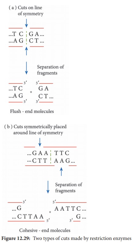
The dashed line is the center of symmetry of the sequence (Table 12.2).
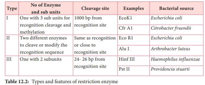
Table 12.2: Types and features of restriction enzyme
Application of Recombinant DNA Technology
a. Production of medically useful proteins such as somato stain, insulin, human growth
hormone and interferon. It decreases the dependency on human tissues and solves
problem of limited production
b. Development of synthetic vaccines for instance,
vaccines for malaria and rabies a recombinant hepatitis vaccine is already
commercially available.
c. Gene therapy
d. Diagnosis of infection diseases.
e. To
manufacture industrially important products like enzymes using bacteria, fungi
and cultured mammalian cells
Related Topics