Chapter: Forensic Medicine: Basic anatomy and physiology
Respiration

Respiration
The respiratory organs
In the preceding discussion we distinguished
between the systemic circulation which nourishes the tissues of the body, and
the respiratory circulation which purifies the blood. The purifying process is
brought about by respiration, or breathing. The organs of respiration are the
air passages and the lungs (fig 2.11, 2.14).
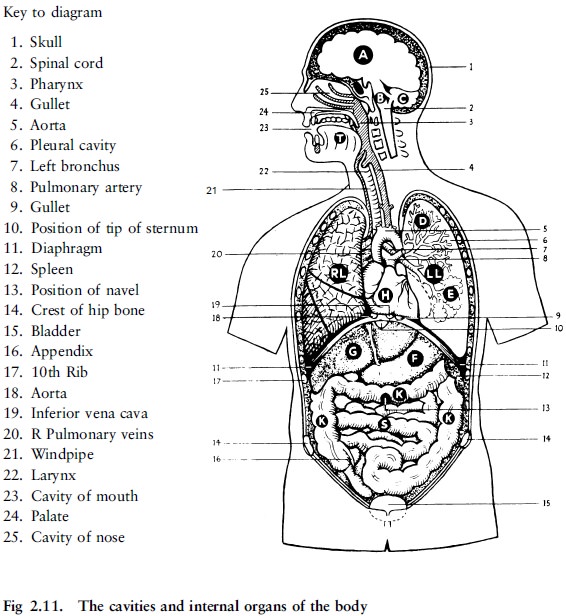
The air passages
The air passages convey the air to the lungs.
They are the following:
·
The nose. When passing through the nasal cavities the air
is warmed, moistened, and freed from dust and germs. The fine hair in the nasal
cavities act as coarse filter, preventing foreign matter from being inhaled.
The nose is also the seat of the sense of smell and can give warning of the
presence of harmful or foul air. However, this sense is not totally reliable as
many poisonous gases have no odour. In quiet breathing the nose is the natural
passage for the air. In deeper breathing the mouth is also used. The cavity of
the mouth has already been described as lying between the upper and lower jaw
bones, which carry the teeth. It is separated from the nasal cavities by the
palate, bony in front (the hard palate), composed of soft parts at the back
(the soft palate). The floor of the mouth is muscular and give attachment to
the tongue. The mouth and the nose both open at the back into the upper part of
the throat or pharynx.
·
The upper part of the throat or pharynx. The tonsils are situated at the point where the
mouth joins the throat, one on either side. From the throat the air passes by
means of the larynx to the windpipe.
·
The larynx. The larynx lies at the upper end of the
windpipe, just below and behind the tongue. It is the organ of voice. Its upper
opening, where the air enters it from the back of the throat, is guarded by a
leaflike valve or lid. During swallowing this lid, which lies over the entrance
of the larynx, prevents food or liquid from entering it. The food, therefore,
passes backwards over the entrance of the larynx, into the gullet, which passes
down the neck behind the windpipe (fig 2.11).
·
The windpipe (trachea). This runs down the vertical middle line of the
neck in front, where the hoops of gristle which protect it and keep it open can
be felt under the skin. It passes into the chest behind the breast bone and,
just above the heart, divides into two main branches.
·
The right and left bronchi. One of these passes to the right and the other
to the left lung.
The lungs
The lungs are the essential organs of breathing.
They lie, one on each side, in the chest cavity and fill the greater part of
this cavity.
The lungs are light, elastic and spongy,
containing air, and very richly supplied with blood. Each is covered by a
smooth membrane (the pleural membrane), and a similar membrane lines the
interior walls of the chest cavity. Between the two membranes there is some
fluid which enables the lungs to move easily during breathing.
After entering the lung the bronchus divides and
subdivides into even smaller branches or air tubes. The very finest of these
tubes end by opening into a tiny space surrounded by a cluster of air sacs that
open into the space into which the air is led in this way (fig 2.21).
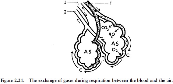
Surrounding each air sac is a close network of
blood capillaries with blood coming from the heart through the pulmonary
artery. The air sacs have very thin walls, and as the blood flows round them,
oxygen from the air in the air sacs passes through their walls, and is taken
into the blood in the capillaries. At the same time carbon dioxide and water
pass out of the blood in the capillaries. In this way, the blood brought to the
lungs by the pulmonary ateries loses carbon dioxide and water, and takes up
oxygen. It thus becomes purified and is then returned to the heart by the
pulmonary veins.
The air in the air sacs at the same gives off
oxygen and takes up carbon dioxide and water, thus becoming impure. When we
breathe out, we expel a certain amount of this impure air; when we breathe in,
we draw in pure air to take its place.
Anything that blocks or stops this exchange of
gases between the blood and the air in the lungs immediately endangers life,
and soon causes death if it is not removed.
Respiration movements
Breathing involves two movements, namely
inspiration or breathing air in, and expiration or breathing air out (fig
2.22).
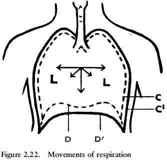
Inspiration happens in two ways: by the action
of muscles which act upon the ribs, thereby raising the walls of the chest so
that its capacity is increased both from front to back and from side to side,
and by the action of the diaphragm, the dome-shaped muscle which separates the
chest from the abdomen. The diaphragm contracts during inspiration, and in
doing so descends and flattens. Thus the capacity of the chest is also
increased from above downwards. The lungs follow the movement of the walls of
the chest and diaphragm, and expand, and thus air is drawn into them. At the
same time the capillaries of the lungs are dilated, so that during inspiration
more blood passes through the lungs.
In ordinary quiet breathing, expiration is only
a slight muscular act. When the effort of inspiration ceases, the walls of the
chest fall back again, the diaphragm relaxes and rises, the elastic lungs
contract, and air is thus driven out of the chest. During exertion, when we
exhale as much air as possible, additional muscular effort is required, as is
the case with inhalation under such circumstances.
Related Topics