Chapter: Forensic Medicine: Basic anatomy and physiology
The blood vessels - Circulation of the blood
The blood vessels
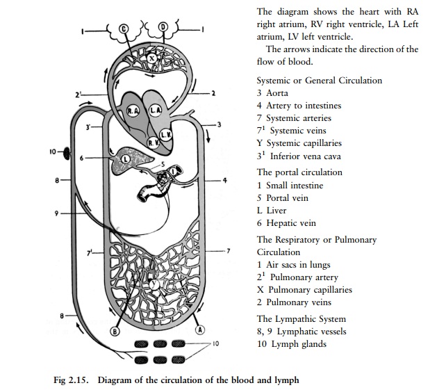
The arteries (fig 2.15, 2.16)
Blood going to all parts of the body from the
heart leaves the left side of the heart through one large vessel, the aorta.
The aorta forms an arch above the heart, and from this arch large branches are
given off on either side, supplying blood to the head and neck (right and left
carotid arteries) and to the upper limbs (right and left subclavian arteries).
The aorta then runs downwards behind the heart alongside the spine, passes
through the diaphragm and enters the abdomen, giving off branches on the way,
mainly to the internal organs of the abdomen, and to the muscles and skin of
the trunk.
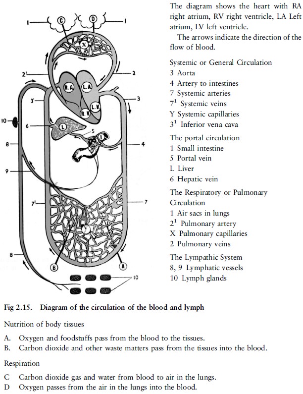
Just below the level of the navel the aorta
divides into two large branches (iliac arteries), one on either side, each of
which goes on ultimately to supply blood to the corresponding lower limb as the
right or left femoral artery.
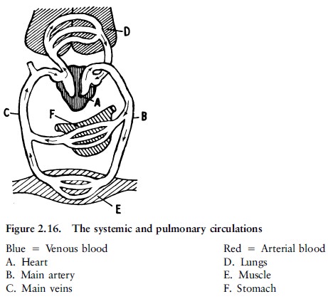
Each of these larger arteries gives off smaller branches, and these again smaller and smaller branches, until finally in the tissues themselves the smallest arteries break up into a network of very fine and short tubes which are called the capillaries and which are so small that they can only be seen through a microscope (fig 2.17).
Capillaries (fig 2,15, 2.17)
It is when blood is passing through the capillaries that the essential work of the blood, namely the nourishment of the tissues is done. The walls of the capillaries are extremely thin and a certain amount of the fluid portion of the blood, containing the nutrients, passes through the thin walls of these very fine tubes and so reaches the tissues. Oxygen also passes from the blood through the walls of the capillaries in a similar way. In this way the tissues are kept supplied both with food and oxygen. Conversely, much of the waste matter formed by the activity of the tissues, such as carbon-dioxide gas, passes from the tissues through the walls of the capillaries into the bloodstream and is thus carried away.
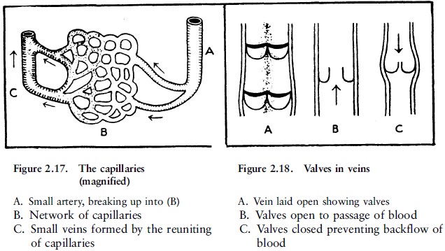
Veins (fig 2.15, 2.18)
The capillaries in turn join together to form
larger tubes which are the small veins, and these again join ever larger veins,
corresponding in size to the arteries and frequently running alongside them. In
this way the veins carry blood back to the heart. Ultimately one large vein
(the superior vena cava) receives all the blood from the upper limbs and from
the head and neck, and another large vein (the inferior vena cava), the blood
from the lower limbs and the abdomen, and both of these large veins enter the
right side of the heart. Most of the veins, especialy those of the limbs have,
all along their length on the inside many small valves which prevent a backflow
of the blood (fig 2.18).
All parts of the body, skin, bones, muscles and
internal organs, are supplied with blood in this way. To each part the blood is
carried from the heart by arteries, through each part it passes in capillaries,
and from each part it is carried away again by veins back to the heart.
The lymph and the lymphatic vessels (fig 2.15)
That part of the fluid portion of the blood
which escapes from the capillaries into the spaces of the tissues, as just
described, is called the lymph. The lymph is not returned immediately into the
blood, but is drained off from the tissues by a separate system of small tubes
or vessels called lymph vessels. These join together just as veins do to form
even larger vessels, although the lymphatic vessels are smaller in size than
the veins. Finally two large lymph branches empty all the lymph back again into
the blood in the great veins at the root of the neck. The lymphatic vessels
thus form a part of the system of organs by means of which the circulation of
the body fluids is maintained. This is shown diagrammatically in figure 2.15.
The lymphatic vessels of the small intestine
have an additional duty namely the absorption of the fats which pass by way of
these vessels to larger lymphatic branches and so to the great veins at the
root of the neck (fig 2.15).
There is one other point of importance regarding
the lymphatic system. On the course of the lymphatics in various parts of the
body small sieve-like structures called lymph glands are interposed (fig 2.14).
These serve as a barrier against the entrance of germs of infection into the
general circulation.
Related Topics