Chapter: Forensic Medicine: Basic anatomy and physiology
The skeleton
The skeleton
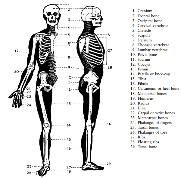
The skeleton (fig 2.1)
The skeleton forms the framework around which
the body is built up and which gives the body its general shape. The bones of
the skeleton give the muscles a place to attach to, and muscle contraction
makes movement possible.
A muscle starts out from one bone and is
inserted into another bone across a joint. When the muscle shortens or
contracts, the joint is moved, permitting movement or locomotion. Bones such as
the skull, the spinal column, the ribs and the hip bones protect some very
important internal organs.
The main purpose of the limbs is to provide a
system of levers to make body movement possible. The limb bones are typically
long bones with strong shafts and large round ends.
On the other hand, bones whose main purpose is
to protect important internal organs are usually flat bones, for example the
bones of the skull, the ribs, the breastbone and the hip bones. Where a part of
the skeleton is designed for compactness, with limited movement, as at the
wrist or the back part of the foot, or in the backbone, the bones are typically
short bones, and are frequently very irregular in shape.
The skeleton is usually divided into three
parts:
·
the central rod, consisting of the skull and spinal column, with the
ribs and breastbone attached to the spine
·
the bones of the upper limb
·
the bones of the lower limb
The Central axial skeleton
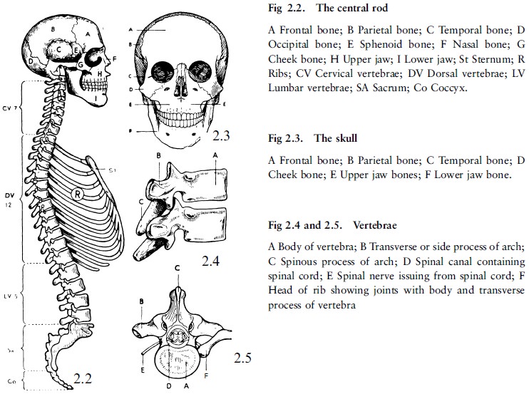
The Skull
The skull consists of two parts.
The brain case or cranium
This forms the upper part of the skull and
encloses and protects the brain.
The cranium consists of eight bones. Its front
is formed by the frontal bone and its back by the occipital bone. Between
these, forming the crown of the head and the upper part of the sides of the
skull, are the two parietal bones, one on either side. Below the parietal bones
are the two temporal bones in which the middle and inner ear are lodged on
either side.
The floor of the cranium is formed by the lower
portion of these bones (except the parietals) with, in addition, in the front
part of the floor, the sphenoid bone and the ethmoid bone which form part of
the bridge of the nose and the eye sockets.
The upper portion of the cranium is smooth and
dome-shaped, and is called the vault. The vault is composed of several flat,
curved, saucer-shaped bones which are firmly joined. The vault as a whole,
however, is sufficiently elastic to give slightly under a blow, thus
diminishing the effects of the blow. This characteristic is more evident in
children, whose skull bones, like the other bones, are more elastic than
adults' bones.
The floor of the cranium is called the base, and
is in line with the eyebrows and the openings of the ears. The base is more
irregular in thickness and more rigid than the vault. The base of the skull
rests on the upper part of the bones of the spine. In the back half of the base
(in the occipital bone) there is a large opening, the foramen magnum, through
which the nerve matter of the brain joins that of the spinal cord. Other
smaller openings in the base give passage to nerves and blood vessels. The
openings leading to the inner ear are easily recognised (in the temporal bone)
on either side.
The bones of the face
The bones of the face form the lower portion of
the skull in front. These bones are firmly joined together, with the exception
of the lower jaw, which is the only movable bone in the skull.
There are 14 bones in the face. Most of them are
arranged in pairs, one on either side of the face. The largest are the upper
and the lower jaw bones, which are the main bones for chewing or mastication.
Some other important bones are the nasal bones, which give shape to the nose,
the cheek bones, which give prominence to the cheeks, and the palate bones,
which separate the cavity of the nose from that of the mouth. The remaining five
bones are small bones, which form the eye sockets and the interior of the nose.
The lower jaw consists of two halves joined
together at the middle line in front, at the chin. Each half is rectangular in
shape with a horizontal portion containing the sockets for the teeth. These
halves run backwards on either side from the front of the chin to beneath and a
little in front of the lower end of the outer ear. Here the lower jaw forms an
angle (the ``jaw'' angle) with the vertical portion, which runs upwards to be
joined to the base of the skull at a depression just in front of the opening of
the ear.
In the upper part of the face on each side are
the orbits or eye sockets, the cavities in which the eyes are lodged. At the
back of each orbit is an opening through which the optic nerve passes back to
the brain from the eye. In the middle of the face are the nose cavities
separated from each other by a narrow partition (septum) running from the back
along the middle line. Between the upper and the lower jaw is the mouth cavity.
The upper and the lower jaws carry the teeth. The cavities of the nose and
mouth are separated from each other by the palate bones.
The spine or vertebral column (26 bones fig 2.2)
The spine consists of 26 separate bones.
Twenty-four of these are similar in shape, and each is called a vertebra. The
lower end of the spine is formed by a large wedge-shaped bone, the sacrum or
rump bone, and beneath this is a small structure, the coccyx, which forms the
tip of the spine.
Generally speaking, each vertebra is built along
the same pattern (fig 2.4 and 2.5). In front it consists of a short solid
cylinder of bone called the body. At the back on either side there are bony
processes which unite at the middle line behind to form the arch. From the arch
at either side, and also at the middle line behind, there are other bony
processes.
The process at the back of the arch is called
the spinous process of the vertebra; the processes on either side of the arch
are called the transverse processes.
The vertebrae are arranged one above the other
in a column. In front the bodies form a column of short solid cylinders which
carry the weight of the trunk and head, and are seen to increase in size and
strength from the neck downwards. Behind, the arches form a column or series of
rings which enclose a canal (the spinal canal) and in which lies the spinal
cord (fig 2.5). Between each pair of vertebrae is an opening on either side
through which a spinal nerve issues from the spinal cord. Throughout the whole
length of the spine the vertebrae are bound together by strong fibrous bands or
ligaments, but between the body of each vertebra and that of the vertebra above
and below it, is a pad or disc of cartilage or gristle (fig 2.4). These discs
are elastic. They allow slight movement between the bodies and act as buffers
against jolts caused by blows on the head or falls where the person lands on
the lower end of the spine or on the feet.
Although the amount of movement possible between
any one pair of vertebrae is small, the sum of these movements in the spine as
a whole allows bending forwards or backwards or to either side, and also to a
lesser extent a twisting of the spine.
There are seven vertebrae in the neck (cervical
vertebrae). The 12 vertebrae in the upper part of the trunk (dorsal vertebrae)
give attachment to the ribs. In the loin area there are five (lumbar
vertebrae). The spine is curved slightly forward in the neck, backward in the
dorsal regions, and forward again in the region of the loins (fig 2.2).
The sacrum or rump bone consists of five
vertebrae, fused together, and much altered in shape, to form one wedge-shaped
bone. It fits between the hip bones behind, transmitting the weight of the body
to them, and so to the lower limbs. The coccyx is a small bone attached to the
lower end of the sacrum, and forms the tip of the spine.
The ribs (12 pairs) and the breastbone (figs 2.1 and 2.2)
The ribs are flat curved bones, 12 on each side.
They are numbered from top to bottom. The ribs are joined at the back to the bodies
and side transverse processes of the dorsal vertebrae (fig 2.5), and arch
forward, enclosing the cavity of the chest. The upper seven are joined by means
of small joints to the breastbone, but between each rib and the breastbone is a
short plate of elastic cartilage or gristle, an arrangement which makes elastic
chest movement possible when breathing. The next three ribs are joined by
similar plates of cartilage to the cartilage of the seventh rib. The lowest two
ribs are shorter and have no attachment in front. They are therefore called
floating ribs.
The breastbone (sternum) is a flat,
dagger-shaped bone lying on the middle line in front. It is joined by small
joints to the two collar bones above, and at either side to the ribs in the way
already described.
The spine at the back, the ribs at the sides,
and the breastbone in front together form the bony portion of the chest (or
thorax) and protect the organs within.
The bones of the upper limb
The shoulder girdle
The bones of the arm proper are joined on either
side to the trunk by two bones, the collar bone in front, and the shoulder
blade behind. The two pairs of bones form an incomplete girdle at the upper
part of the trunk, which is called the shoulder girdle.
The collar bone (or clavicle) is a strong
double-curved bone running directly outwards, from its joint with the upper end
of the breastbone, to the point of the shoulder, where it is joined by a small
joint to a process of bone which springs from the back of the shoulder blade.
The shoulder blade (or scapula) is a flat,
triangular bone which lies at the back of the shoulder. A process of the
shoulder blade is joined to the outer end of the collar bone to form the point
of the shoulder. The shoulder joint lies below the point of the shoulder, and
is formed by the bone of the upper arm and the shoulder blade.
The rounded head of the bone of the upper arm
fits into a shallow socket on the outer edge of the body of the shoulder blade,
thus forming the joint (fig 2.9). The shoulder blade does not join any bone
behind, but is attached to the ribs and backbone by muscles.
The bones of the arm
The bones of the arm proper are the bone of the
upper arm (1); the bones of the forearm (2); the bones of the wrist (8); the
bones of the hand (5); the bones of the thumb and fingers (14).
The bone of the upper arm (or humerus) is a long
bone running from the shoulder joint above, where the head of the bone is
joined to the shoulder blade, to the elbow joint below, where its expanded
lower end is joined to the two bones of the forearm. The shaft of the bone is
compact and strong.
The bones of the forearm are called the radius
and the ulna. The radius is on the outer side, opposite the thumb, the ulna on
the inner side, opposite the little finger. They run from the elbow joint to
the wrist, where they form the wrist joint with the upper row of wrist bones.
There are also joints between the radius and the ulna, which allow hand-turning
so that the hand can be turned palm up or with the back of the hand up. In this
movement the ulna remains stationary and the radius turns round it.
There are eight wrist bones (carpal bones),
forming two rows, an upper and lower, of four each. They are small irregularly
shaped short bones. There are small joints between them which allow a limited
amount of movement between the bones.
There are five hand bones (metacarpal bones)
which are joined at the top to the lower row of the wrist bones, and at the
bottom to the thumb and fingers. They have the characteristic shape of long
bones. Each of the fingers has three small long bones (phalanges); the thumb
has only two.
The bones of the lower limb
The hip girdle
The hip bones are two large bones, one on either
side of the body, each shaped roughly like a twin-bladed screw. They are firmly
joined to the sacrum behind, and meet together in front, so that, along with
the sacrum, they form a complete girdle called the pelvis or pelvic girdle.
They transmit the mass of the trunk to the thigh
bones. The rounded head of each thigh bone fits into a deep socket on the outer
side of the corresponding hip bone, thus forming the hip joint (fig 2.7).
The lower half of the pelvis encloses and gives
protection to the bladder, the lower end of the bowel and to other organs.
The bones of the lower limb proper
The bones of the lower limb proper are: the
thigh bone (1); the bones of the leg (two) and the kneecap (one); the bones of
the back part of the foot (7); the bones of the fore-part of the foot (5); the
bones of the toes (14).
The thigh bone (or femur) is the largest and
strongest bone in the body. It is a long bone and runs from the hip joint
above, where it is joined to the hip bone, to the knee joint below. The rounded
head of the femur, which forms part of the hip joint, is joined at an angle, by
means of a short neck, to the straight shaft of the bone. The lower end of the
femur is enlarged forming a prominence on the inner and outer side immediately
above the knee joint (fig 2.8).
Three bones enter into the knee joint: the
femur, the tibia or shin bone, and the kneecap, a small, roughly triangular
bone, placed in front of the joint to strengthen it.
The bones of the lower leg are the shin bone (or tibia), the sharp edge of which may be felt under
the skin at the front of the leg, and the splint bone (or fibula), a slighter
bone, which lies alongside the tibia and is joined to it on its outer side.
Only the inner of the two, the tibia, enters
into the knee joint above, but the lower ends of the both enter into the ankle
joint below, where they join the ankle bone (or talus) which is the uppermost
of the bones of the foot. The lower ends of the tibia and the fibula form the
prominences of the ankle on the inner and outer side respectively.
The bones of the back of the foot and heel (tarsal bones) are short bones. They correspond
to the wrist bones in the upper limb, but are much larger and stronger (they
carry the weight of the body). They number seven. The largest and strongest is
the heel bone (or calcaneum), on which the ankle bone rests.
There are five bones in the fore-part of the
foot (metatarsal bones); each opposite a toe.
The bones of the back and the front of the foot
together form a double arch at the instep, an arrangement which gives a certain
spring to the walk and reduces the effects of jars in jumping or running.
The bones of the toes (phalanges) are arranged
like those of the fingers - two
for the big toe, and three for the others. All the living bones, especially the
long bones, have great power of repair. Consequently, when a bone has been
broken, it is capable of becoming firmly joined together again by the formation
of new bone between and around the broken ends. This process of bone union of a
broken bone takes many days, and in the case of larger bones often a good many
weeks to complete. During the first part of that time the broken bones must be
kept totally immobile.
The joints
Varieties of joints
A joint is a structure which unites two or more
bones, and commonly allows movement between them. The joints are divided into
three main groups in accordance with their degree of movement.
·
joints so firmly joined together that no movement can occur between
them, for example the bones of the cranium
·
joints that allow a limited amount of movement, for example joints
between the vertebrae, between the bones at the back of the foot, or the two
joints between the skull (occipital bone) and the first vertebra of the neck
where the nodding movements of the head take place
·
joints that move freely, such as the joints between the long bones of
the limbs. There are two main types:
a.
the hinge joint where there is backwards and forwards movement on one
level only, as in an ordinary hinge. The elbow (fig 2.9), the joints of the
fingers and toes and, broadly speaking, the knee (fig 2.6) and the ankle are of
this type.
b.
the ball and socket joint, or universal joint, which allows free
movement in all directions. Two joints, the hip (fig 2.7) and the shoulder (fig
2.9) are of this type. The socket of the shoulder joint is shallow and for this
reason accidental dislocation or displacement of the bone of the upper arm at
this joint is not uncommon.
Although these are the more important types, there are also other types of joints. The wrist joint is an example. It has a shallow oval socket or cup (the lower end of the radius and ulna) and a shallow and oval head (the upper row of wrist bones). The range of movement is not as free as that at the shoulder or hip, but much freer than the movement in a simple hinge joint. The movement of turning the head from side to side is performed at a ring and pivot joint between the first and second vertebrae of the neck, the pivot being formed by an upright peg of bone attached to the upper surface of the body of the second vertebra, the first vertebra forming an incomplete ring round the front and sides of the peg. The ring is completed behind by a fibrous ligament.
Structure of a joint (figs 2.6, 2.7 and 2.9)
In the structure of a typical freely movable
joint, the following features are to be noted:
·
The ends of the bones entering into it are enlarged, rounded and smooth,
and shaped to fit on top of each other.
·
Where the bones come into contact they are capped by a thin plate of
smooth cartilage or gristle which allows movement with the least possible
amount of friction.
·
A fibrous capsule or sheath connects the ends of the bones together all
round the edges of the joint, thus enclosing a cavity - the joint cavity. The inner surface of this
capsule is lined by a membrane (the synovial membrane), which secretes a clear
sticky fluid to lubricate the joint - synovial fluid or joint fluid.
·
Lastly, the bones forming the joint are bound together by strong fibrous
bands called ligaments. These are usually found outside the joint, and can be
specially strengthened parts of the capsule. In joints which are subject to
great strain, for example the knee and hip joints, there are special ligaments
inside the joint also. These ligaments support the joint and limit its range of
movement.
The skeleton: medico-legal applications
In general the bones of the body are formed from
gristle (cartilage). The conversion of this cartilage into bone occurs by a
process known asossification, whereby lime salts are deposited in the
cartilage, which is then converted into the characteristic structure of bone.
Ossification begins in all people at almost the same places known as centres of
ossification. Finding these ossification centres assists in age determination
before birth: the centre of ossification of the heel bone (calcaneus) is for
example present by the end of the fifth month of foetal life. At the end of the
seventh month the centre of ossification has appeared in one of the ankle bones
(the talus). By the end of the ninth month or just before birth there is a
well-developed centre at the lower end of the femur or thigh bone.
Information about the presence of these centres
of ossification therefore assists in determining whether an infant has reached
full term or whether it is legally viable (the presence of the ossification
centre in the talus indicating a foetal age of seven months).
In the case of the skull cap, that is the top
and the sides of the skull, the bones are pre-formed not in gristle but in a
tissue called membrane, which is converted into bone. The sutures or lines of
closure between these bones fuse together at different ages. This makes it
possible to determine a person's age, for example by radiological examination
in the living, and by an inspection of the skull itself, when this is the only
part of the skeleton available. This estimate is only an approximate one.
Certain features of the skeleton, for example
the shape of the pelvic cavity, its inlet and its outlet, are determined by the
sex of the subject. There are also certain sex differences in the bones which
make up the pelvis. It is possible therefore to obtain a certain amount of
information from the skeleton which can assist in sex determination when only
skeletal remains are available.
A person's probable height can be determined
from skeletal remains if at least one of the long bones, for example the femur,
is available. The humerus is less reliable than the femur. Other long bones are
not sufficiently reliable for such deductions.
From the skull bones certain deductions can be
made about race which can assist in a limited way in identifying or
distinguishing racial types.
Many of the bones have a central cavity filled
with bone marrow. In the adult the marrow contains a considerable amount of
fat. When bones are broken the fat cells are disrupted and can be forced into
the blood stream. The fat particles can reach vital parts of the brain and
obstruct their blood supply. This can cause death and is known as death from
fat embolism.
An embolus consists of something foreign to the
blood stream. It may be a portion of blood clot, a collection of air, or
particles of fat which gain entrance to the blood stream and which may lodge in
or obstruct blood vessels, thus cutting off the blood supply to the affected
part.
When a fracture involves the surfaces of a
joint, arthritis may develop afterwards.
Related Topics