Chapter: Surgical Pathology Dissection : The Female Genital System
Pelvic Exenterations Including Vaginectomies

Pelvic Exenterations Including Vaginectomies
Vaginectomies
for vaginal cancer include a por-tion of vagina attached to the uterus and
cervix. These specimens can be handled in the same manner as radical
hysterectomies for cervical cancer, although the paracervical soft tissues may
not be present. Note that a clinical history of pre-natal diethylstilbestrol
(DES) exposure is related to the presence of vaginal adenosis and clear cell
adenocarcinoma of the vagina and cervix. Aden-osis appears as a red, granular
change on the normally smooth, white vaginal mucosa. Also look for structural
abnormalities of the cervix and fallopian tubes associated with DES exposure.
Important observations include the size of the tumor and the distance of the
tumor to the vaginal margin. If the uterus has been previously re-moved, the
resulting vaginal pouch can be opened along one side and handled in the same
manner as a large skin excision. Sections should be taken so as to demonstrate
the greatest depth of tumor invasion, the tumor with adjacent normal-appearing
mucosa, and the relationship of the tumor to the cervix. If the bladder is
included with the uterus the resection is termed an anteriorexenteration, and if the rectum is included theresection is
termed a posterior exenteration. With
these added structures, additional sections in-clude documentation of the
extent of tumor in-volvement of the bladder or rectal wall, and an evaluation
of their respective surgical margins. Specifically, these include the urethral
and ure-teral margins for the bladder, and the proximal and distal bowel
margins for the rectum.
Exenterations
are also performed for centrally recurrent cervical cancer. Perhaps the most
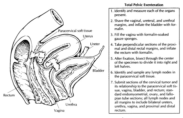
daunting specimen received in the surgical pa-thology laboratory is a total
pelvic exenteration, which includes the bladder, uterus with attached adnexa,
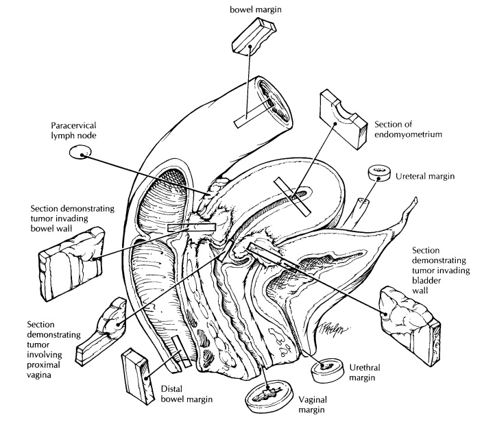
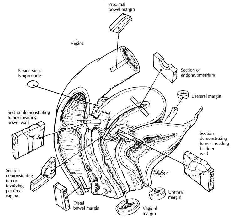
vagina, and rectum. The evaluation of these specimens uses both a separate and
an integrated approach, as described. Resection margins are best handled if
each of the four main components (i.e., bladder, vagina, uterus, and rectum) is
thought of separately. Appropriate examination of the central tumor involves
demonstrating its in situ
relationship to these surrounding organs.![]()
When a
total pelvic exenteration specimen is received for recurrent cervical cancer,
do not panic. Instead, calmly note the organs present and their dimensions.
Specifically, look for the ureters, urethra, bladder, uterus, fallopian tubes,
ovaries, vagina, and rectum. Take shave sections of the vaginal, ureteral, and
urethral margins. Take perpendicular sections from the proximal and distal
rectal margins, providing ink for margin orientation. Next, ink all the exposed
soft tissue that surrounds the cervix and tumor.
Fill the
vagina with formalin-soaked gauze pads, and distend the bladder and rectum with
formalin. Submerge the entire specimen in forma-lin, and fix it overnight. The
fixed specimen may then be bisected in a sagittal plane to demonstrate the
tumor and its relationship to surrounding structures. This is best accomplished
by using probes in the urethra and uterine canal as midline guides. After the
specimen has been sectioned, a diagram can facilitate the description of the
tumor, including its extension. Take sections of the tumor to demonstrate
invasion of the bladder, rectum, vagina, and/or paracervical tissue. Docu-ment
the vaginal and paracervical soft tissue mar-gins with perpendicular or shave
sections. Last, dissect the soft tissue surrounding the cervix, and submit for
histology a section of any lymph nodes found.


Important Issues to Address in Your Surgical Pathology Report on Pelvic Exenterations
· What
procedure was performed, and what structures/organs are present?
· What is
the site of origin of the tumor?
· What are
the histologic type and grade of the tumor?
· What is
the size of the tumor?
· What
other organs are involved by the tumor? Specify the extent of tumor involvement
into these structures. That is, does it reach the mus-cular wall, submucosa, or
mucosa?
· Does the
tumor infiltrate the capillary–lym-phatic spaces?
· Does the
tumor involve any resection margins? Give the distance of the tumor from the
closest margin (in centimeters).
· Does the
tumor involve any lymph nodes? In-clude the number of nodes involved and the
number of nodes examined at each specified site.
· Are any
radiation effects present?
Related Topics