Chapter: Essentials of Anatomy and Physiology: Reproductive System
Oogenesis and Fertilization - Female Reproductive System
Oogenesis and Fertilization
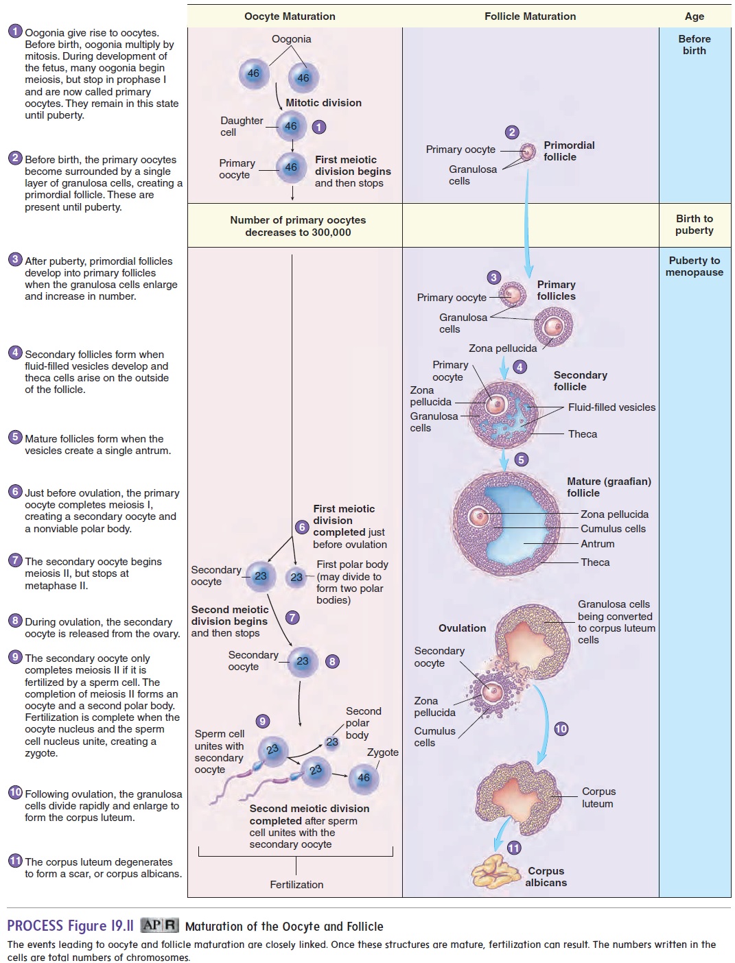
Ovulation is the release of an oocyte from an ovary (figure 19.11,step 8). Just before ovulation, the primary oocyte completes the first meiotic division to produce a secondary oocyte and a polar body. Unlike meiosis in males, cytoplasm is not split evenly between the two cells. Most of the cytoplasm of the primary oocyte remains with the secondary oocyte. The polar body either degenerates or divides to form two polar bodies. The secondary oocyte begins the second meiotic division but stops in metaphase II.
The zygote has 23 pairs of chromosomes (a total of 46 chromosomes). All cells of the human body contain 23 pairs of chromosomes, except for the male and female gametes. The zygote divides by mitosis to form 2 cells, which divide to form 4 cells, and so on. The mass of cells formed may eventually implant in, or attach to, the uterine wall and develop into a new individual .
Follicle Development
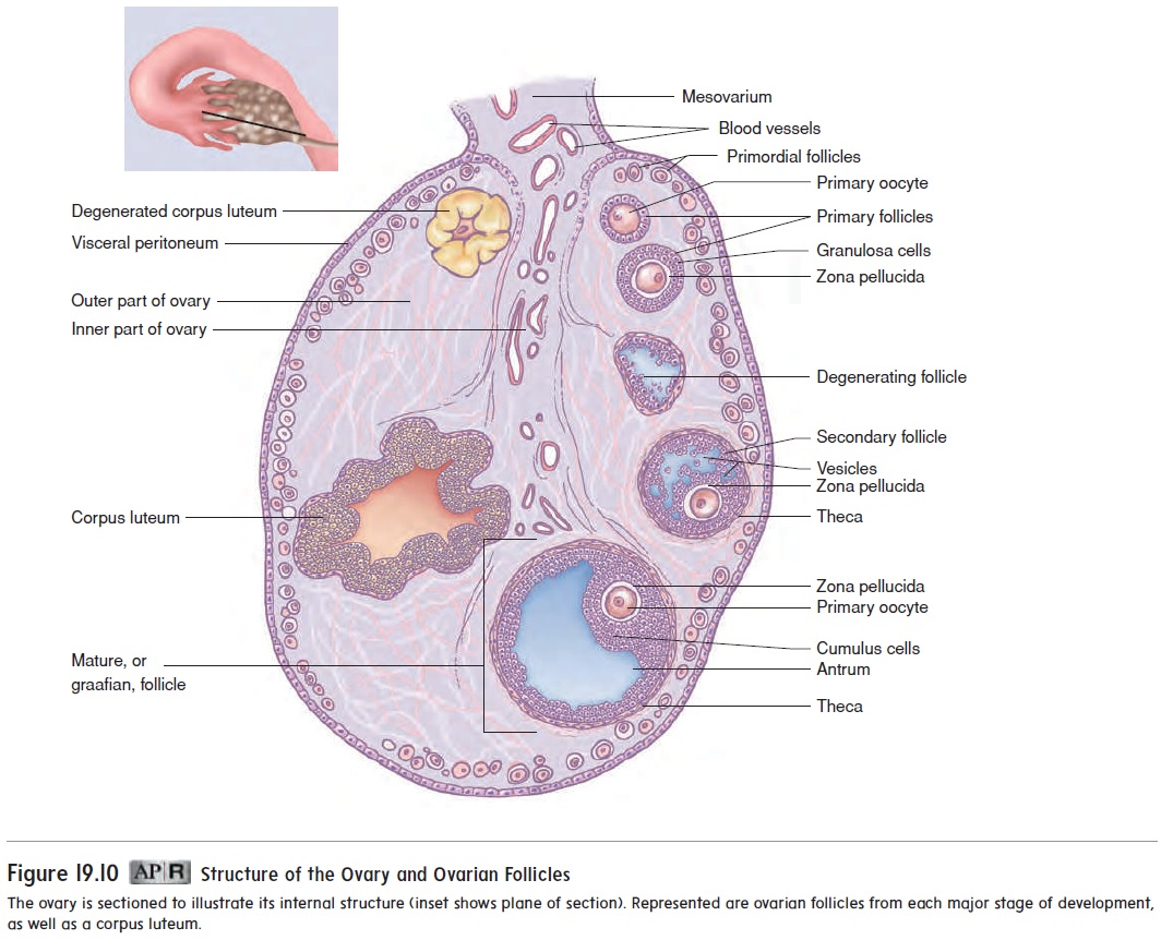
As we discussed, when a female is in her mother’s uterus, her ovaries have already begun oocyte formation. The primary oocytes present at birth are surrounded by a primordial follicle. A primordial follicle is a primary oocyte surrounded by a single layer of flat cells, called granulosa cells (figure 19.11). Once puberty begins, some of the pri-mordial follicles are converted to primary follicles when the oocyte enlarges and the single layer of granulosa cells becomes enlarged and cuboidal. Subsequently, several layers of granulosa cells form, and a layer of clear material called the zona pellucida (z̄o ′ n̆a pel-l̄u ′ sid-d̆a ) is deposited around the primary oocyte.
Approximately every 28 days, hormonal changes stimulate some of the primary follicles to continue to develop (figure 19.11). The primary follicle becomes a secondary follicle as fluid-filled spaces called vesicles form among the granulosa cells, and a cap-sule called the theca (thē ′ kă ; a box) forms around the follicle.
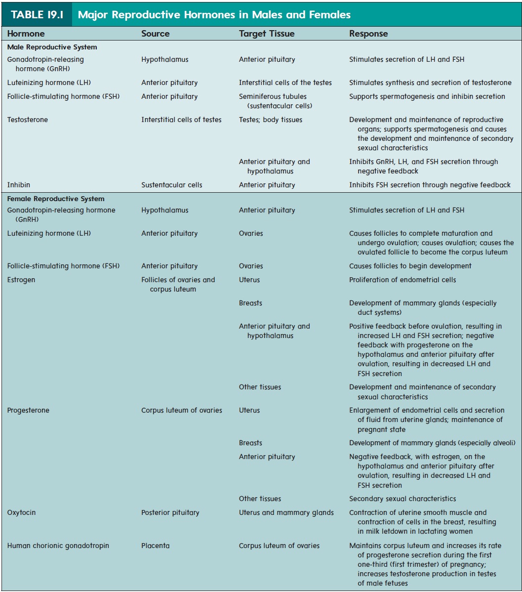
The mature follicle forms a lump on the surface of the ovary. During ovulation, the mature follicle ruptures, forcing a small amount of blood, follicular fluid, and the secondary oocyte, surround-ed by the cumulus cells, into the peritoneal cavity. In most cases, only one of the follicles that begin to develop forms a mature follicle and undergoes ovulation. The other follicles degenerate. After ovu-lation, the remaining cells of the ruptured follicle are transformed into a glandular structure called the corpus luteum (k̄o r′ p̆u s, body; loo′ t̄e -̆u m, yellow). If pregnancy occurs, the corpus luteum enlarges in response to a hormone secreted by the placenta called humanchorionic gonadotropin hormone (hCG) (k̄o-r̄e-on′ik ḡo′nad-o-tr̄o ′ pin) (see table 19.1). If pregnancy does not occur, the corpus luteum lasts for 10–12 days and then begins to degenerate.
Related Topics