Chapter: Microbiology and Immunology: Virology, Virus: Arboviruses
Mosquito-Borne Flaviviruses
Mosquito-Borne Flaviviruses
◗ Encephalitis viruses
St. Louis encephalitis virus: St. Louis encephalitis virus is the most important mosquito-borne viral disease in the United States. It is transmitted by Culex mosquitoes. Wild birds, such as English sparrows, are the reservoir hosts. Humans are the dead-end host. These viruses are mostly seen in urban areas because these mosquitoes breed in stagnant waste water. The virus causes encephalitis, which is associated with a mortality rate of 10%. Isolation of the virus is difficult; hence diagnosis is mostly based on serology. No antiviral therapy or vaccine is available.
Ilheus virus: The virus occurs mostly in South and CentralAmerica. The infection is maintained in forests by bats and mosquitoes. The virus usually causes asymptomatic infection and rarely causes encephalitis.
West Nile virus: West Nile virus causes encephalitis. The virusis endemic in the Middle East, Africa, and Asia. Wild birds are the main reservoirs of the virus. The infection is transmitted from wild birds to humans by Culex, Aedes, and Anopheles mos-quitoes. Humans are the dead-end hosts. The virus first causes asymptomatic or symptomatic illness in the wild migratory birds. These infected birds contain high titer of the viruses and remain viremic for 1–2 weeks. These serve as important reser-voirs of infection for transmission to humans, horses, dogs, and other small animals after being bitten by mosquitoes.
The virus like other arthropod-borne viruses causes vire-mia, crosses blood–brain barrier, and infects the brain paren-chyma causing viral encephalitis. Patients with this virus may present with features of encephalitis, aseptic meningitis, or both. This condition has also been reported from India, and the virus has been isolated from Culex mosquitoes. The virus has been isolated from the brain of these fatal cases of encephalitis in Karnataka in 1980–1981. Mental confusion and disorientation with decreased consciousness are some of the manifestations of patients with encephalitis. The infec-tion usually occurs in summer months when humans, mos-quitoes, and wild migratory birds remain in close proximity outdoors.
Laboratory diagnosis of the condition is made by isolation of the virus from blood within the first 2 weeks of infection; the virus cannot be isolated from CSF. Enzyme immunoas-say and plaque reduction utilization test are available for the specific diagnosis of the condition by demonstration of specific antibodies in serum. A fourfold or greater rise of antibody titers between acute and convalescent titer is diag-nostic of the infection. Polymerase chain reaction (PCR) is also available. No antiviral therapy or vaccine for the virus is available.
This is an infection that can be transmitted by transfusion of contaminated blood. Hence, PCR and nucleic acid probes specific for the virus are used to screen the blood for the virus.
Murray Valley encephalitis virus: This virus is endemic inNew Guinea and Australia. The virus occurs in enzootic cycle involving mosquitoes and wild birds. Culex annulirostris is the main vector. The virus was first reported during an epidemic of encephalitis in Murray River Valley in 1951, and hence was called Murray Valley encephalitis virus.
Japanese encephalitis virus: JE virus is a flavivirus, whichcauses JE, the most common cause of epidemic encephalitis. JE virus is a single-stranded RNA virus closely related to St. Louis encephalitis and West Nile virus. The virus was first isolated in Japan during an epidemic of encephalitis in 1935. The virus initially was called Japanese B encephalitis to distin-guish it from the Japanese A encephalitis virus (Wang Ecunoms type A encephalitis), which had different epidemiological prop-erties. In India, the virus was first reported in 1955, during an outbreak of encephalitis in Tamil Nadu.
Birds and pigs are two main reservoir hosts. JE is transmit-ted to humans by certain species of Culex mosquitoes widely prevalent in rice fields in Asia. The disease is spread throughout mostly in rural areas of Asia by culicine mosquitoes, most often Culex tritaeniorhynchus. Natural infection occurs in Ardeid birds,such as herons and egrets, and the virus is transmitted from bird to bird by C. tritaeniorhynchus.
The bite of infected mosquitoes transmits JE virus to humans. The virus initially replicates at the site of the bite and in the regional lymph nodes. Subsequently, the virus spreads through circulation, causing viremia and leading to inflammatory changes in the reticuloendothelial system, heart, lungs, and liver. Usually, most infections are cleared before the virus infects the central nervous system (CNS) causing subclinical disease. However, neurological invasion can also occur possibly by growth of the virus across vascular endothelial cells, thereby affecting the large parts of brain including the thalamus, basal ganglia, brain stem, cerebellum, hippocampus, and cerebral cortex.
Circulating antibodies play an important role in conferring heterologous immunity against JE.
Most of the infections are asymptomatic. Only 1 in 250 infections results in symptomatic disease. Most symptomatic infections occur in young children between 2 and 10 years and in elderly people in endemic areas. In nonendemic areas, the infection occurs in all the age groups.
Incubation period of JE varies from 4 to 15 days. The condi-tion has an abrupt onset with appearance of symptoms, which include fever, headache, nausea, diarrhea, vomiting, and myal-gia, which may last for several days. This is followed by altered state of consciousness, tremors, incoordination, and convul-sions. Seizures develop in two-thirds of people—most often in children, while headache and meningismus are more common in adults.
Mortality rate is high; in less-developed areas with poor medical care, the mortality rate may exceed 35%. More than 10,000 reported deaths occur per year due to JE worldwide. The neurological sequelae are severe and can occur in many patients who suffer from the disease. The neurological sequelae include seizure disorder, movement disorder, or motor or cranial nerve paralysis.
JE is a seasonal disease with most cases occurring in temper-ate areas from June to September. In subtropical areas, trans-mission begins as early as March and extends until October. Transmission may occur earlier in some subtropical areas, such as Indonesia. Approximately, 35,000–50,000 symptom-atic cases are reported per year worldwide, although this is slightly an underestimation of the true incidence of the disease. Approximately 3 billion people currently live in areas endemic for JE, which extend from India to Maritime, Siberia, and Japan.
The disease occurs throughout Asia, but is most prevalent in Southeast Asia.
JE in India was first reported from Vellore, Tamil Nadu, and Pondicherry in 1950s. Since then the condition has been reported in all the states and union territories of India. An estimated 378 million people are living at the risk of JE in 12 states, union territories that are frequently affected. Several outbreaks of JE have occurred in Bankura and Burdwan in West Bengal, Dibrugarh in Assam, South Arcot in Tamil Nadu, Kolar in Karnataka, various areas in Andhra Pradesh, and in Pondicherry, and Gorakhpur in Uttar Pradesh. Rapid agricultural development and intensive rice cultivation sup-ported by irrigation systems facilitating breeding of culicine mosquitoes is primarily responsible for the spread of the condition to newer areas in India. An epidemic of JE virus occurred in Uttar Pradesh and Bihar in India in 2005 and also throughout Nepal. During this epidemic, more than 5000 cases were reported and approximately 1000 deaths were documented.
C. tritaeniorhynchus is the most important mosquito spe-cies that transmits JE. This mosquito has a peculiar habit to bite outdoors and is extremely active in the evening and night. These mosquitoes breed in collection of water, typically rice fields, thereby increasing the risk of infections in rural areas. Low-grade, short-term viremia occurs in humans and other mammals; hence these are dead-end hosts for the virus. Pigs are the amplifying hosts, because they have persistent, high-grade viremia and help in transmission of the virus in humans living close to the pig-rearing areas.
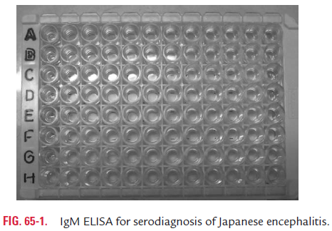
Laboratory diagnosis of JE is made by isolating the virus from the blood during the first week of illness. The CSF rarely yields virus except in severe or fatal cases. Serodiagnosis is made by using ELISA for demonstration of IgM antibodies in serum or CSF (Fig. 65-1). This test has a high sensitivity, nearing 100%. The test may show false-positive reaction with serum from other febrile illnesses, such as dengue and West Nile fever or with the serum collected after vaccination with yellow fever and JE. The IgM antibodies are present in serum or CSF in approximately three-fourths of patients within the first 4 days after onset of illness and in nearly all the patients after 7 days of illness. Direct fluorescent antibody test is a rapid method for detection of viral antigen directly in the brain tissue. Reverse transcriptase polymerase chain reaction (RT-PCR) is used to demonstrate viral genome in the CSF and blood for diagnosis of JE.
There is no specific antiviral therapy available for treatment of JE. Prevention consists of the use of inactivated mouse-brain-derived vaccine using the Nakayama strain. Mosquito eradication and measures to prevent mosquito bite, such as use of mosquito nets, mosquito repellants, and insecticides are useful to prevent transmission of infection.
◗ Yellow fever virus
Yellow fever is a mosquito-borne acute febrile illness that occurs in Africa and South America. The disease is not reported in India. Yellow fever virus is the type-specific virus of the family Flaviviridae. It is a single-stranded, enveloped, RNA virus. The envelope consists of a lipid bilayer containing an envelope gly-coprotein and a matrix protein. The single RNA is complexed with a capsid protein.
Yellow fever is transmitted by the bite of Ades mosquito. During blood meal, the mosquito deposits the saliva contain-ing the virus into a bite wound. The virus replicates locally and in regional lymph nodes draining the wound. Subsequently, the virus spreads by blood to the bone marrow, liver, myocardium, spleen where further replication of virus occurs. The condition is associated with hemorrhagic manifestations occurring as a result of disseminated intravascular coagulation. This occurs due to bleeding from the gastrointestinal mucosa and from abdominal and pleural serous layers. This is associated with reduction in the synthesis of coagulation factor and altered platelet function. The condition progresses to shock and finally death due to mul-tiple organ failure involving liver, kidney, brain, and heart.
Host immunity is characterized by the presence of viral neu-tralizing antibodies by the end of the first week during which the virus is rapidly cleared from the circulation. The role of immunity in the pathogenesis of the disease is not known. An attack of yellow fever confers a lifelong immunity.
Incubation period is short and varies from 3 to 6 days. No prodromal symptoms occur.
As the name suggests, yellow fever is characterized by jaun-dice and fever. This is an illness characterized by an acute onset of fever followed by jaundice within 2 weeks of onset of symp-toms. Typically, symptoms begin suddenly with fever, chill, malaise, prostration, headache, giddiness, myalgia, anorexia, nausea, and vomiting. This may be associated with bleed-ing from the nose, gums, gastrointestinal tract, or skin that usually occurs within a few weeks of illness. Hemorrhagic dia-thesis progressing to disseminated intravascular coagulation is the most important complication of the disease. Multiple organ failure involving liver, kidney, and heart is responsi-ble for death of the patient. Yellow fever is a severe and life-threatening disease.
The yellow fever is endemic in South America and in the Caribbean enzootic countries of Africa. Although yellow fever can be transmitted in Asian countries, till now no documented transmission of yellow fever has been reported from Asian coun-tries including India. It has been suggested that previous infec-tion with another flavivirus, such as dengue virus, may confer protection from yellow fever virus. This is the reason suggested for the absence of yellow fever in Asian countries including India. Yellow fever shows following transmission cycles:
Jungle (sylvatic) cycle: In this cycle, the mosquito transmitsthe virus to wild, nonhuman primates; from these hosts, it is transmitted to another mosquito. Humans are the incidental hosts in the cycle. This sylvatic cycle is present in rain forests, and the humans, such as men clearing the trees in the forest are bitten by the mosquito. Aedes species are the main vectors in Africa. Wild Aedes species are the vectors in South America.
Urban cycle: This cycle is confined to urban areas in whichthe mosquitoes transmit the virus to a human host and then it is transmitted to another mosquito. A. aegypti, a domestic mos-quito, is the primary vector.
Intermediate cycle: This cycle is confined to Savanna forestarea of Africa and is the most common cycle responsible for outbreaks of yellow fever in Africa. In this cycle, the mosquito transmits the virus to wild nonhuman primates and human hosts and then it is transmitted to another mosquito.
The yellow fever is transmitted from infected humans to mosquitoes, which are diurnal feeders. Humans suffering from the disease during initial 3–4 weeks of illness are the source of infection. The extrinsic incubation period is 12–21 days. Vertical transmission of yellow fever takes place from female mosquitoes to the female progeny in 1% of cases. This is the important reason for the survival of the virus in dry season. Approximately, 3–10 variants are necessary to infect a mos-quito. Seasonal transmission of yellow fever occurs during mid-rainy season and early dry season in Africa. In South America, this occurs from January to March. An estimated 200,000 cases of yellow fever occur in Africa and South America, causing 20,000–30,000 deaths annually. Laboratory diagnosis of yellow fever can be made by:
· Isolation of yellow fever virus from the clinical specimens.
· Demonstration of IgM-specific antibodies in the serum or demonstration of fourfold or more rise in serum IgG.
· Detection of yellow fever antigen in tissues by immunohistochemistry
· Detection of viral genomes by PCR, and
· Elevated transaminase and bilirubin levels, which are dem-onstrated during the toxic stage of illness.
Vaccination is the most widely used preventive measure against yellow fever. Other preven-tive measures include mosquito eradication program and personal protective measures. The latter include the use of proper clothings, insect repellants, etc., which avoid being bitten by mosquitoes and thereby exposure to yellow fever virus.
◗ Dengue virus
Dengue is the most common mosquito-borne arboviral ill-ness caused by dengue virus. The name dengue is derived from a Swahili word ki-dinga-pepo meaning a sudden seizure by a Demon. The earliest known documentation of symptoms of dengue-like illness was described in Chinese Encyclopedia dur-ing 265 AD. In 1780, Rauss coined the term “break-bone fever” based on description of symptoms reported by patients during Philadelphia epidemics of probably dengue fever. The possible outbreak of dengue fever epidemics were documented sporadi-cally every 10–30 years until World War II. Subsequently, after World War II the dengue fever spread and became worldwide. The first epidemic of dengue hemorrhagic fever was described in 1963 in Manila. In 1979–1980, the first reported outbreak of dengue fever occurred simultaneously in Asia, Africa, and North America.
Related Topics