Chapter: Microbiology and Immunology: Morphology and Physiology of Bacteria
Microscopy and Types of Microscopy
Microscopy
Microscopy is an important component of diagnostic micro-biology. Bacteria being very small cannot be visualized by the naked eye, because the limit of resolution with the unaided eye is about 200 microns. So, the study of bacteria requires the use of microscopes. Amicroscope is an instrument that uses one or more lenses to produce a magnified image of an object that is invisible to the unaided eye.
Types of Microscopy
The following types of microscopy are used for the examination of microorganisms including bacteria:
◗ Light microscopy
Light microscopy, as the name suggests, uses natural or artificial transmitted light as the source of light. Resolving power of microscope is an important component of light microscopy. It is the ability of the lens system to distinguish two closely placed objects as distinct and separate entities. It is dependent on the wavelength of light used to illuminate the object and on the numerical aperture of the microscope. It is about half of the wavelength of light being used. For example, the smallest particle which can be resolved by yellow light with a wavelength of 0.4 mm is about 0.2 mm. Proper use of condenser that focuses light on the plane of the object facilitates optimization of the resolving power of the microscope. Resolving power of the microscope is enhanced further by adjusting the medium through which light passes between the object and objective lens. The use of immersion oil, whose refractive index is similar to that of the glass, improves the resolution of the microscope. The numerical aperture of the microscope is defined as the light gathering power of the microscope. Different types of light microscopy include (a) bright-field microscopy, (b) dark-ground microscopy, (c) phase-contrast microscopy, and (d) interference microscopy.
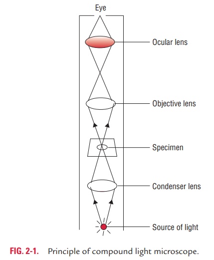
1. Bright-field microscopy: Bright-field microscopy (alwaysreferred to as ordinary light microscopy) is the most common form of light microscopy that uses a compound light micro-scope. A compound light microscope primarily consists of a compound lens system that contains a number of objective lenses, such as lenses of low power (310), high power (340), and oil immersion (3100). It also contains a fixed ocular (eye piece) lens, usually of 310 or 35. Final magnification of an object is the multiplication of lens power of the objective with that of the eye piece (Fig. 2-1). The bright-field microscopy has many uses.
It may be used to examine either wet films or “hanging drop” for demonstration of the motility of flagellated bacteria (e.g.,Escherichia coli, Pseudomonas aeruginosa, etc.) and protozoa (e.g., Trichomonasvaginalis, Giardia intestinalis, etc.). The wet prep-aration is also useful for demonstration of microorganisms in urine or feces, and also for detection of fungi in the skin.It is useful for demonstration of the structural details.
It is also useful for measuring approximate size of the bacte-ria, fungi, and protozoa in stained preparations.
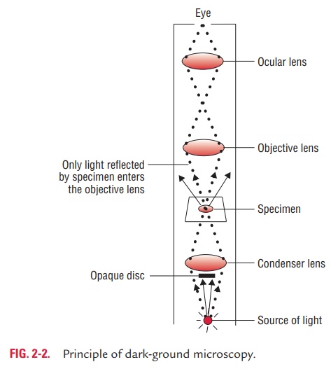
2. Dark-ground microscopy: The dark-ground microscopymakes use of dark-ground microscope, a special type of compound light microscope. The dark-field condenser with a central circular stop, which illuminates the object with a cone of light, is the most essential part of the dark-ground microscope. This microscope uses reflected light instead of transmitted light used in the ordinary light microscope (Fig. 2-2). It prevents light from falling directly on the objective lens. Light rays falling on the object are reflected or scattered onto the objective lens with the result that the microorganisms
· It is useful for demonstration of very thin bacteria (such as, spirochetes) not visible under ordinary illumination, since the reflection of the light makes them appear larger. This is a frequently used method for rapid demonstration of Treponemapallidumin clinical specimens.
· It is also useful for demonstration of motility of flagellated bacteria and protozoa.
3. Phase-contrast microscopy: Phase-contrast microscopymakes use of a specific optical system that converts differences
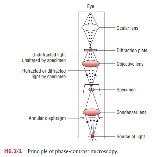
in phase in an organism into differences in intensity of light thereby producing light and dark contrast in the image (Fig. 2-3).
The optical system includes a special condenser and objec-tive lens which can be fitted to an ordinary light microscope to convert it into a phase-contrast microscope. The phase-con-trast microscopy has following uses:
o It is immensely useful for examination of living micro-organisms particularly protozoa (e.g., T. vaginalis, Entamoebahistolytica, etc.)
o It is useful for examining the internal structures of a living cell by improving the contrast and differentiating structures within the cell that differs in their thickness and refractive index.
4. Interference microscopy: This is another specialized appli-cation of light microscopy used for demonstrating cell organ-elles. It is also useful for quantitative measurement of the chemical constituents of the cells, such as proteins, lipids, and nucleic acids.
◗ Fluorescence microscopy
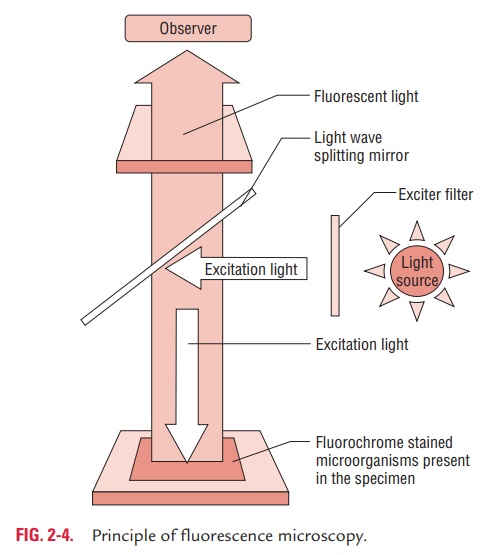
Fluorescence microscopy is based on the principle that the spec-imens stained with fluorescent dye when exposed to ultraviolet light result in emission of longer wavelength of light (i.e., vis-ible light) (Fig. 2-4). The bacteria stained with fluorescent dye appear as a brightly glowing object against a dark background.
Fluorescence microscopy needs a fluorescence microscope fitted with an ultraviolet light source. Auramine O, acridine orange, and rhodamine are fluorescent dyes used to visualize bacteria. The resolving power of a fluorescence microscope is increased due to the short wavelength of ultraviolet light. Auramine O, acridine orange, and rhodamine are fluorescent dyes used to visualize bacteria. Fluorescence microscopy is widely used in diagnostic microbiology in the following ways:
· It is used for direct demonstration of antigen of a patho-gen in clinical specimens by direct fluorescent antibody test (e.g., direct detection of Neisseria gonorrhoeae, Corynebacteriumdiphtheriae, etc. directly in clinical specimens).
· It is also used for the estimation of antibodies in the serum by indirect fluorescent antibody test (IFA) (e.g., IFA in lepto-spirosis, syphilis, brucellosis, etc.).
◗ Electron microscopy
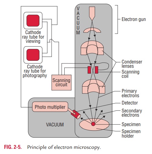
Electron microscopy utilizes a beam of electrons instead of a beam of light used in the light microscopy. The electron beam is focused by electromagnets, analogous to the lenses used in the light microscopy. The object to be examined is kept on the path of the beam that scatters the electrons and produces an image which is focused on a screen (Fig. 2-5).
The resolving power of the electron microscope is extremely high, theoretically 100,000 times than that of a light micro-scope. This is because the electron microscope uses electrons whose wavelength is approximately 0.005 nm as compared to 5000 nm wavelength of the visible light. As mentioned earlier, the resolving power is half of the wavelength. In practice, the resolving power of the electron microscope, however, is about 0.1 nm. There have been many developments in electron microscopy that include (a) shadow casting, (b) scanning elec-tron microscopy, (c) immunoelectron microscopy, and (d) freeze-etching, etc.
Shadow casting is an important technique that is carried outby depositing a thin layer of platinum or other metal on the microorganism to be examined. This platinum-coated organism, on bombardment with electron beams, scatters the electron and produces an image that is focused on a fluorescent screen.
Scanning electron microscopy is another development thatprovides a three-dimensional image of the object as well as high resolution.
Immunoelectron microscopy is a method to enhance sensi-tivity and specificity by reacting the specimen with specific antiviral antibody that results in clumping of viral particles. In this method also, antibody may be conjugated with gold to visualize and determine the location of specific antigenic determinants in a specimen.
Freeze-etching is the method by which live organisms can be visualized unlike in traditional methods of electron microscopy in which living cells cannot be examined. This method is use-ful for the study of cellular ultrastructure of the microorgan-isms in the living state. This method is based on rapid cooling of specimens by deep-freezing in liquid gas and the subsequent formation of carbon platinum replica of the specimen.
Electron microscope is widely used for:
· Rapid detection of viruses directly in clinical specimens. This is especially useful for detection of noncultivable viruses.
· Ultrastructural study of various microorganisms.
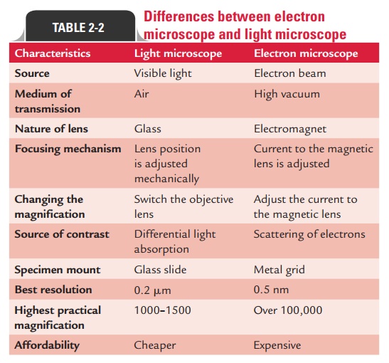
Differences between electron microscope and light microscope are summarized in Table 2-2.
◗Newer microscopic methods
These include the following:
Confocal microscopy: This is useful to obtain highresolution images and for three dimensional reconstruction of biological models.
Scanning probe microscopy: This measures surface fea-tures by moving a sharp probe over the object’s surface. There are two types of scanning probe microscope: (a) scan-ning tunneling microscope and (b) atomic force microscope.
Related Topics