Chapter: Microbiology and Immunology: Morphology and Physiology of Bacteria
Electron microscopy
Electron microscopy
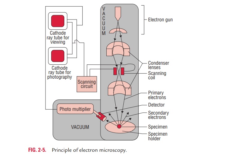
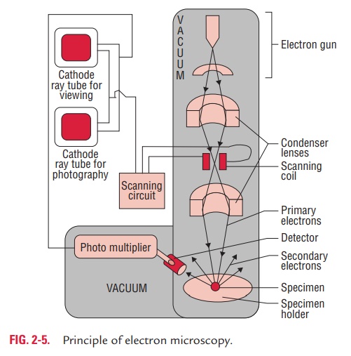
Electron microscopy utilizes a beam of electrons instead of a beam of light used in the light microscopy. The electron beam is focused by electromagnets, analogous to the lenses used in the light microscopy. The object to be examined is kept on the path of the beam that scatters the electrons and produces an image which is focused on a screen (Fig. 2-5).
The resolving power of the electron microscope is extremely high, theoretically 100,000 times than that of a light micro-scope. This is because the electron microscope uses electrons whose wavelength is approximately 0.005 nm as compared to 5000 nm wavelength of the visible light. As mentioned earlier, the resolving power is half of the wavelength. In practice, the resolving power of the electron microscope, however, is about 0.1 nm. There have been many developments in electron microscopy that include (a) shadow casting, (b) scanning elec-tron microscopy, (c) immunoelectron microscopy, and (d) freeze-etching, etc.
Shadow casting is an important technique that is carried outby depositing a thin layer of platinum or other metal on the microorganism to be examined. This platinum-coated organism, on bombardment with electron beams, scatters the electron and produces an image that is focused on a fluorescent screen.
Scanning electron microscopy is another development thatprovides a three-dimensional image of the object as well as high resolution.
Immunoelectron microscopy is a method to enhance sensi-tivity and specificity by reacting the specimen with specific antiviral antibody that results in clumping of viral particles. In this method also, antibody may be conjugated with gold to visualize and determine the location of specific antigenic determinants in a specimen.
Freeze-etching is the method by which live organisms can be visualized unlike in traditional methods of electron microscopy in which living cells cannot be examined. This method is use-ful for the study of cellular ultrastructure of the microorgan-isms in the living state. This method is based on rapid cooling of specimens by deep-freezing in liquid gas and the subsequent formation of carbon platinum replica of the specimen.
Electron microscope is widely used for:
· Rapid detection of viruses directly in clinical specimens. This is especially useful for detection of noncultivable viruses.
· Ultrastructural study of various microorganisms.
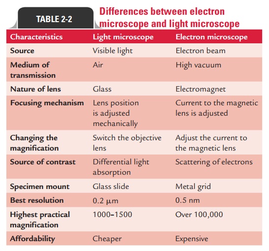
Differences between electron microscope and light microscope are summarized in Table 2-2.
Related Topics