Chapter: BIOLOGY (ZOOLOGY) Standard XI first year 11th text book Assignment topics question and answer Explanation Definition
Compound light, Dark field, Phase contrast, Oil - immersion and Electron microscope
Compound light, Dark field, Phase contrast, Oil - immersion and Electron microscope
Compound light microscope
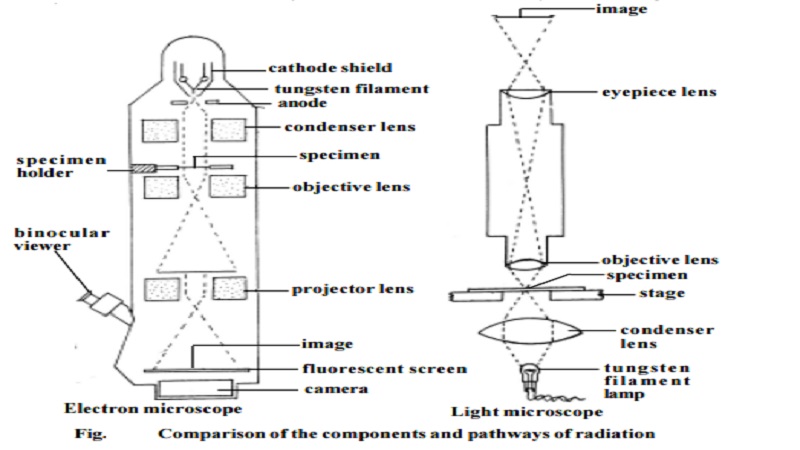
This microscope uses visible light for illuminating the object. It contains glass lenses that magnify the image of the object and focus the light on the retina of the observer's eye. It has two lenses one at each end of a hollow tube. The lens closer to the object being viewed is called objective lens. The lens closer to the eye is called ocular lens or eyepiece. The object is illuminated by light beneath it. A third lens called condenser lens is located between the object and the light source and it serve to focus the light on the object.
Dark field microscope
This type of microscope is useful for viewing suspensions of bacteria. It has a special condenser that allows only rays of light scattered by struc-tures within specimen. The result is an image that appears bright against a dark background, with a high degree of contrast. The process is similar to seeing dust particles floating in a sunbeam.
Phase contrast microscope
The phase contrast microscope has special fitments to the objective lens and sub stage condenser, the effect of which is to exaggerate the structural differences between the cell components. As a consequence, the structures within living, unstained cells become visible in high contrast and with good resolution. Phase contrast microscopy avoids the need to kill cells or to add dye to a specimen before it is observed microscopically.
Oil - immersion microscopy
In oil-immersion microscopy the light gathering properties of the ob-jective lens are enhanced by placing oil in the space between the slide and objective lens. Normally the technique is used to view permanently mounted specimens.
The oil immersion lens gives higher magnification than the normal high-power objective lens.
Electron microscopy.
The electron microscopy uses the much shorter wavelengths of elec-trons to achieve resolutions as low as 3�. Electromagnetic coils (ie., mag-netic lenses) are used to control and focus a beam of electrons accelerated from a heated metal wire by high voltages, in the range of 20,000 to 100,000 volts. The electrons are made to pass through the specimen. Electrons that do passes out of the object are focused by an objective coil ('lens'). Finally a magnified image is produced by a projector coil or 'lens'. The final image is viewed on a screen or is recorded on photographic film to produce electron micrograph. This type of electron microscope is called transmission electron microscope (TEM)
In a compound light microscope, the image is formed due to differences in light absorption. The electron microscope forms images as a result of differences in the way electrons are scattered by various regions of the object.
The degree to which electrons are scattered is determined by the thick-ness and atomic density of the object. Hence the specimens used in electron microscopy must be extremely thin. Living cells which are wet cannot be viewed in electron microscope.
Scanning electron microscopy (SEM)
This microscope has less resolution power than the TEM (ie., about 200�). However it is a very effective tool to study the surface topography of a specimen. The whole specimen is scanned by a beam of electrons. An im-age is created by the electrons reflected from the surface of the specimen. Scanning electron micrographs show depth of focus and a three dimensional image of the
Related Topics