Chapter: Medicine and surgery: Respiratory system
Coal worker’s pneumoconiosis - Occupational lung disease
Coal worker’s pneumoconiosis
Definition
Pathology resulting from inhalation of coal dust and its associated impurities.
Prevalence
Two per 1000 coal workers.
Aetiology
The disease is caused by dust particles approximately 2–5 µm in diameter that are retained in the small airways and alveoli of the lung.
Pathophysiology
Two different syndromes result from inhalation:
Simple pneumoconiosis in which there is deposition of coal dust within the lung. There are peribronchiolar deposits in the upper parts of the lung, often associated with mild centriacinar emphysema.
Progressive massive fibrosis (PMF): The pathogenesis is not understood. Patients develop rheumatoid and antinuclear factor and the damage is thought to be due to immune complexes.
Clinical features
Simple pneumoconiosis is asymptomatic. Patients with progressive massive fibrosis suffer from considerable effort dyspnoea, usually with a cough. The sputum may be black.
Macroscopy/microscopy
· Simple pneumoconiosis is characterised by accumulation of dust in macrophages at the centre of the acinus, with associated emphysema.
· In progressive massive fibrosis there are nodules of >3 cm in the upper lobes. Histologically the nodules can be divided into three types:
i. Amorphous collection of acellular proteinaceous material, containing little collagen and abundant carbon, which frequently cavitates and liquefies. Seen where silica content is low.
ii. Dense collagenous tissue and macrophages heavily pigmented by carbon, seen where there is a high silica content in the coal dust.
iii. Caplan’s syndrome seen where there is co-existent rheumatoid disease. Carbon-stained rheumatoid nodules are seen.
Complications
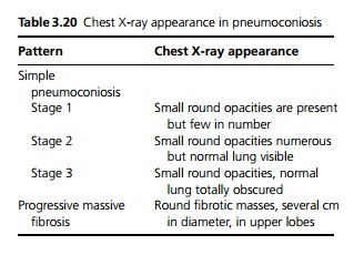
Simple pneumoconiosis is divided into three stages by chest X-ray appearance (see Table 3.20). Stage 1 does not progress, 7% of patients with stage 2 and 30% of patients with stage 3 will go on to develop progressive massive fibrosis. PMF by definition is progressive, and respiratory failure will eventually develop.

Investigations
The diagnosis is made by chest X-ray in those who have been exposed (see Table 3.20).
Related Topics