Chapter: Psychiatric Mental Health Nursing : Neurobiologic Theories and Psychopharmacology
Brain Imaging Techniques
BRAIN IMAGING TECHNIQUES
At one time, the brain could be studied only through surgery or
autopsy. During the past 25 years, however, several brain imaging techniques
have been developed that now allow visualization of the brainŌĆÖs structure and
function. These techniques are useful for diagnosing some disorders of the
brain and have helped to correlate certain areas of the brain with specific
functions. Brain imaging techniques are also useful in research to find the
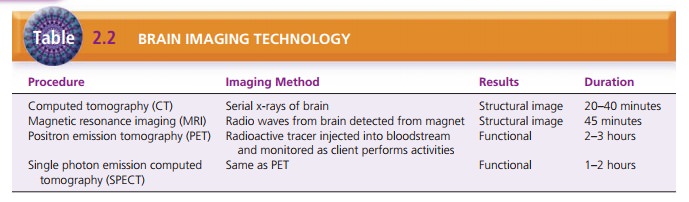
causes of mentaldisorders. Table 2.2 describes and compares several of these
diagnostic techniques.
Types of Brain Imaging Techniques
Computed tomography (CT), also called computed axial tomography (CAT), is a procedure in
which a precise x-ray beam takes cross-sectional images (slices) layer by
layer. A computer reconstructs the images on a monitor and also stores the
images on magnetic tape or film. CT can visualize the brainŌĆÖs soft tissues, so
it is used to diagnose primary tumors, metastases, and effusions and to
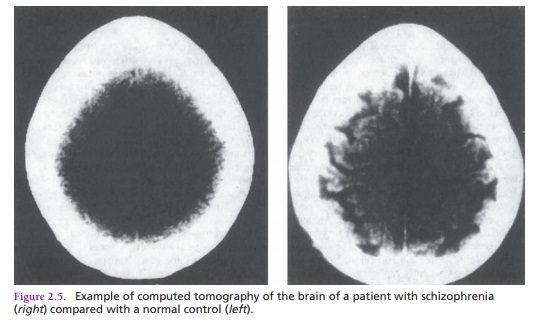
determine the size of the ventricles of the brain. Some people with
schizophre-nia have been shown to have enlarged ventricles; this finding is
associated with a poorer prognosis and marked negative symptoms (Figure 2.5;)
The person under-going CT must lie motionless on a stretcher-like table for
about 20 to 40 minutes as the stretcher passes through a tunnel-like ŌĆ£ringŌĆØ
while the serial x-rays are taken.
In magnetic resonance imaging
(MRI), a type of body scan, an energy field is created with a huge magnet
andradio waves. The energy field is converted to a visual image or scan. MRI
produces more tissue detail and contrast than CT and can show blood flow
patterns and tissue changes such as edema. It also can be used to measure the
size and thickness of brain structures; persons with schizophrenia can have as
much as 7% reduction in cortical thickness. The person undergoing an MRI must
lie in a small, closed chamber and remain motionless during the procedure,
which takes about 45 minutes. Those who feel claustro-phobic or have increased
anxiety may require sedation before the procedure. Clients with pacemakers or
metal implants, such as heart valves or orthopedic devices, can-not undergo
MRI.
More advanced imaging techniques, such as positron emission tomography
(PET) and single photon emission
computed tomography (SPECT), are used to examine the function of the brain. Radioactive substances are injected into
the blood; the flow of those substances in the brain is monitored as the client
performs cognitive activities as instructed by the operator. PET uses two
photons simul-taneously; SPECT uses a single photon. PET provides better
resolution with sharper and clearer pictures and takes about 2 to 3 hours;
SPECT takes 1 to 2 hours. PET and SPECT are used primarily for research, not
for the diagnosis and treatment of clients with mental disorders (Fujita,
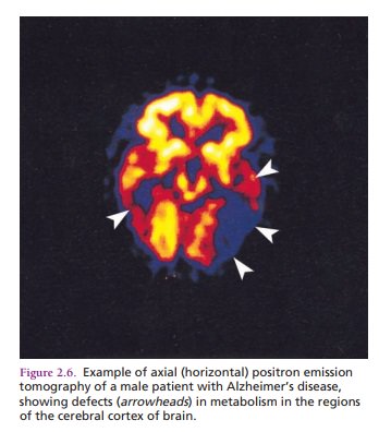
Kugaya, & Innis, 2005; Vythilingam et al., 2005) (Figure 2.6). A recent breakthrough
is the use of the chemical marker FDDNP with PET to identify the amy-loid
plaques and tangles of AlzheimerŌĆÖs disease in living clients; these conditions
previously could be diagnosed only through autopsy. These scans have shown that
cli-ents with AlzheimerŌĆÖs disease have decreased glucose metabolism in the
brain and decreased cerebral blood flow. Some persons with schizophrenia also
demonstrate decreased cerebral blood flow.
Limitations of Brain Imaging Techniques
┬Ę
Although imaging techniques such as PET and SPECT have helped bring
about tremendous advances in the study of brain diseases, they have some
limitations:
┬Ę
The use of radioactive substances in PET and SPECT limits the
number of times a person can undergo these tests. There is the risk that the
client will have an aller-gic reaction to the substances. Some clients may find
receiving intravenous doses of radioactive material frightening or
unacceptable.
┬Ę
Imaging equipment is expensive to purchase and maintain, so
availability can be limited. A PET camera costs about $2.5 million; a SPECT
camera costs about $500,000.
┬Ę
Some persons cannot tolerate these procedures because of fear or
claustrophobia.
┬Ę
Researchers are finding that many of the changes in dis-orders such
as schizophrenia are at the molecular and chemical levels and cannot be
detected with current imaging techniques (Fujita et al., 2005; Vythilingam et
al., 2005).
Related Topics