Chapter: Human Neuroanatomy(Fundamental and Clinical): Blood Supply of Central Nervous System
Blood Supply of the Spinal Cord - Blood Supply of Central Nervous System

Blood Supply of Central Nervous System
The nervous system is richly supplied with blood. Interruption of blood supply even for a short period can result in damage to nervous tissue. Traditionally it has been taught that lymphatics are not present in nervous tissue, but some workers have recently challenged this view.
Blood Supply of the Spinal Cord
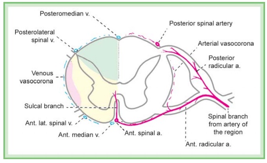
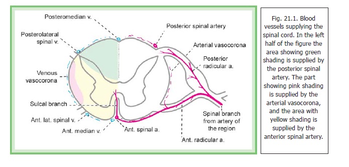
The spinal cord receives its blood supply from three longitudinal arterial channels that extend along the length of the spinal cord (Fig. 21.1). Theanterior spinal artery is present in relation to the anterior median fissure. Two posterior spinal arteries (one on each side) run along the posterolateral sulcus (i.e., along the line of attachment of the dorsal nerve roots). In addition to these channels the pia mater covering the spinal cord has an arterial plexus (called the arterialvasocorona) which also sends branches into the substance of the cord.
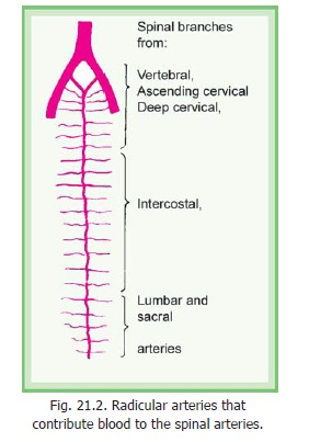
The main source of blood to the spinal arteries is from the vertebral arteries (from which the anterior and posterior spinal arteries take origin). However, the blood from the vertebral arteries reaches only up to the cervical segments of the cord. Lower down the spinal arteries receive blood through radicular arteries that reach the cord along the roots of spinal nerves. These radicular arteries arise from spinal branches of the vertebral, ascending cervical, deep cervical, intercostal, lumbar and sacral arteries (Fig. 21.2).

Many of these radicular arteries are small and end by supplying the nerve roots. A few of them, which are larger, join the spinal arteries and contribute blood to them. Frequently, one of the anterior radicular branches is very large and is called the arteria radicularis magna. Its position is variable. This artery may be responsible for supplying blood to as much as the lower two-thirds of the spinal cord.
The greater part of the cross sectional area of the spinal cord is supplied by branches of the anterior spinal artery (Fig. 21.1, left half). These branches enter the anterior median fissure (or sulcus) and are, therefore, called sulcal branches. Alternate sulcal branches pass to the right and left sides. They supply the anterior and lateral grey columns and the central grey matter. They also

supply the anterior and lateral funiculi. The rest of the spinal cord is supplied by the posterior spinal arteries. As already mentioned branches from the arterial vasocorona also supply the cord.
The veins draining the spinal cord are arranged in the form of six longitudinal channels. These are anteromedian and posteromedian channels that lie in the midline; andanterolateral and posterolateral channelsthat are paired (Fig. 21.1). These channels are interconnected by a plexus of veins that form avenous vasocorona. The blood from theseveins is drained into radicular veins that open into a venous plexus lying between the duramater and the bony vertebral canal (epidural or internal vertebral venous plexus) andthrough it into various segmental veins.
Clinical:
Thrombosis in the anterior spinal artery produces a characteristic syndrome. The territory of supply includes the corticospinal tracts. This leads to an upper motor neuron paralysis below the level of lesion. The spinothalamic tracts are also involved. This leads to loss of sensations of pain and temperature below the level of lesion. Touch and conscious proprioceptive sensations are not affected as the posterior column tracts are not involved. The extent of damage varies depending on the efficiency of anastomoses in the region.
Related Topics