Locomotion and Movement | Zoology - Answer the following questions | 11th Zoology : Chapter 9 : Locomotion and Movement
Chapter: 11th Zoology : Chapter 9 : Locomotion and Movement
Answer the following questions
Zoology
Locomotion and Movement
Evaluation
Answer the following questions
21. Name the different types of movement
1.
Amoeboid movement
2.
Cilliary movement
3.
Flagellar movement
4.
Muscular movement
22. Name the filaments present in the sarcomere
1.
Thick filament
2.
Thin filaments
23. Name the contractile proteins present in the skeletal muscle
1.
Myosin - Thick filament
2.
Actin - Thin filament
24. When describing a skeletal muscle, what does “striated” mean?
• The striations in the skeletal muscle
mean's the dark a band s and the light I band.
25. How does an isotonic contraction take place?
• In isotonic contraction the length
of the muscle changes but the tension remains constant.
(eg)
lifting dumbbells and weight lifting
26. How does an isometric contraction take place?
• In isometric contraction the length
of the muscle does not change but the tension of the muscle changes, (eg) Pushing
againsta wall holding a heavy bag.
27. Name the bones of the skull.
• The cranial bones are 8 in numbers.
1)
Paired parietal
2)
Paired temporal
3)
frontal
4)
Sphenoid
5)
Occipital
6)
Ethmoid
28. Which is the only jointless bone in human body?
• The joint less bone is hyoid bone
in our throat. The hyoid bone (lingual bone) is a horse shoe shaped bone
situated in the anterior mid line of the neck between the chin and the thyroid
cartilage.
29. List the three main parts of the axial skeleton
1.
Cranium
2.
Hyoid (Lingual)
3.Vertebral
column
4.
Thorasic cavity.
30. How is tetany caused?
• Due to the deficiency of parathyroid
hormone the level of calcium decreases in the blood that leads to rapid muscle spasm
called tetany.
31. How is rigor mortis happened?
• After the death of an individual the
muscle be in a contractile position due to the depletion of ATP that is digested
by lysosomal enzyme called rigor mortis.
32. What are the different types of rib bones that form the rib cage?
1.
True rib bones (First 7 pairs)
2.
False rib bones (8,9,10th pairs)
3.
Floating rib bones (11 and 12th pair)
33. What are the bones that make the pelvic girdle?
1.
Ilium
2.
Ischium
3.
Pubis
34. List the disorders of the muscular system.
1.
Muscle fatique
2.
Atrophy
3.
Muscle pull
4.
Muscular dystrophy
5.
Arthritis a) Osteoarthritis b) Rheumatoid arthritis c) Gout
35. Explain the sliding- filament theory of muscle contraction.
• Sliding filament theory is a active
process. It is proposed by Andrw F. Huxley in 1954 and Rolf Niedergerke.

• Muscle contraction is initiated by
a nerve impluse sents by the central nervous system through a motor neuron
• When the nerve impulse reaches a neuromuscular
junction acetylcholine is released and created action potential.
• This action potential triggers the
release of calcium from sarcoplasmic reticulum
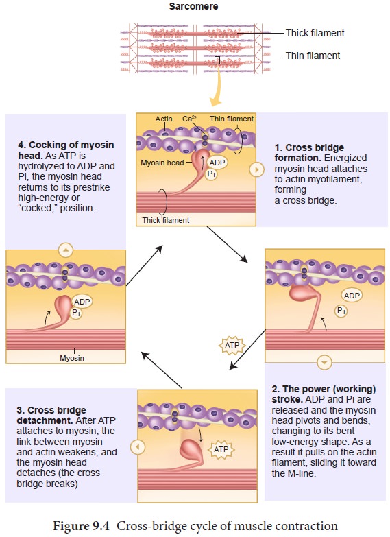
• The released calcium ions binds to
troponin on thin filaments.
• The active sites are exposed to the
heads of myosin to form a cross bridge. Hence actin and myosin form a protein complex
called actomyosin.
• Utilizing the energy released from
hydrolysis of ATP the myosin head rotates until it forms a 90° angle with long axis
of the filament.
• The power stroke begins after the
myosin head and hinge region tilt from a 90° angle to 45° angle.
• The cross bridge transforms into strong
high force bond which allows the myosin head to swellsit
• When the myosin head swells it pulls
the attached actin filament towards the centre of the A - band.
• The myosin returns back to its relaxed state and releases ADP and phosphate ion. A New ATP molecule binds to the head of myosin and the cross bridge is broken.
• At the end of each power stroke each
myosin head detaches from actin then swivels back and binds to a new actin molecule
to start another contraction cycle.
• The power stroke repeats many times
and the thin filaments move toward the centre of the sarcomere.
• In this process there is no change
in the lengths of thick or thin filaments.
• The Z - discs attached to the actin
filaments are also pulled inwards from both the sides causing the shortening of
the sarcomere. This process continues.
• When motor impulse stops the calcium
ions are pumbed back into the sarcoplasmic reticulum results in the masking of the
active sites of the actin filament and the myosin head fails to bind with the actin
and causes Z - discs back to their original relaxed position.
36. What are the benefits of regular exercise?
•
The muscles used in exercise grow larger and stronger.
•
The resting heart rate goes down.
•
More enzymes are synthesized in the muscle fibre.
•
Ligaments and tendons become stronger.
•
Joints become more flexible.
•
Protection from heart attack.
•
Influences hormonal activity.
•
Improves cognitive functions.
•
Prevents Obesity.
•
Promotes confidence, esteem.
•
Aesthetically better with good physique.
•
Over all well- being with good quality of life.
•
Prevents depression, stress and anxiety.
37. What are the different types of bone fracture?
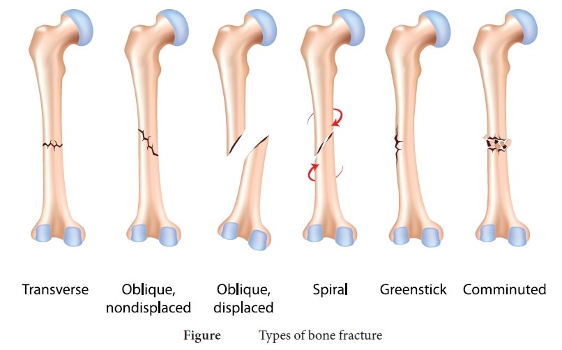
These
are the common types of fractures.

Types
of bone fracture
•
Tranverse - A fracture that is at right angle to the bone's long axis.
•
Oblique non-displaced- A fracture that is diagonal to the bone's long
axis and the fractured bone is not displaced from its position.
•
Oblique displaced - A fracture that is diagonal to the bone's long axis
and the fractured bone is displaced from its position.
•
Spiral - Ragged break occurs when excessive twisting forces are applied
to a bone (common sports fracture).
•
Greenstick - Bone breaks incompletely, just like a green twig breaks. It
is common in children, because of the flexibility of the bones.
•
Comminuted - Bone fragmented into three or more pieces. Particularly
common in the aged, whose bones are brittle (hard but easily broken).
38. Write about the mechanism and healing of bone fracture.
Mechanism
and healing of a bone fracture
•
Bone is a cellular, living tissue capable of growth, self-repair and remodeling
in response to physical stresses. In the adult skeleton, bone deposit and bone
resorption occur. These two processes together constitute in remodeling of
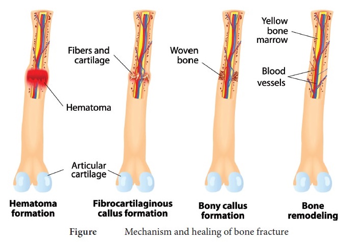
bone. There are four major stages in repairing a simple fracture.

Mechanism and healing of bone
fracture
Formation
of haematoma:
•
When a bone breaks the blood vessels in the bone and surrounding tissues are
torn and results in haemorrhage.
•
Due to this a haematoma, a mass of clotted blood forms at the fracture site.
•
The tissues at the site becomes swollen, painful and inflammed.
•
The death of bone cells, occur due to lack of nutrition.
Formation
of fibrocartilaginous callus :
•
Within a few days several events lead to the formation of soft granulation
tissue called callus.
•
Capillaries grow into the haematoma and phagocytic cells invade the area and
begin to clean up the debris.
•
Meanwhile the fibroblasts and osteoblasts invade from the nearby periosteum and
endosteum and begin reconstructing of the bone.
•
The fibroblasts produce fibres.
•
The chondroblasts secrete the cartilage matrix.
•
Within this repair tissue, osteoblasts begin forming spongy bone.
•
The cartilage matrix later calcifies and forms the fibrocartilaginous callus.
Formation
of Bony callus :
•
New bone trabeculae begin to appear in the fibro cartilaginous callus.
•
Gradually that is converted into a bony - (hard) callus of spongy bone.
•
Bony callus formation continues until a firm union is formed about two months
later to an year for complete woven bone formation.
Remodeling
of Bone Bony callus formation
•
Will be continued for several months.
•
After that the bony callus is remodelled.
•
The excess material on the diaphysis exterior and within the medullary cavity
is removed and the compact bone is laid down to reconstruct the shaft walls.
•
The final structure of the remodelled area resembles like the unbroken bony
region.
39. What is meant by physiotherapy?
•
Physiotherapy is the therapeutic exercise to make the limbs work near normally.
•
It is a rehabilitation profession with a presence in all health care centres.
•
Therapeutic exercises are carried out by physiotherapists.
•
The common problem at the end of fracture treatment is the wasting of muscles
and stiffness of joints.
•
These problems can be restored by the physiotherapy with gradual exercises.
•
It has proven to be effective in the post surgery treatment and management of
arthritis, spondylosis, musculo skeletal disorders, stroke and spinal cord injury.
40. Comment on the dislocation of joints.
Dislocation
of joint is the total displacement of the articular end of the bone from the
joint cavity. The normal alignment of the bones becomes altered. Joints of the
jaw, shoulders, fingers and thumbs are most commonly dislocated. Dislocations
of joints are classified as (i) Congenital deformities (ii) Traumatic (iii)
Pathological (iv) Paralytic.
•
Congenital deformities are due to genetic factors or factors operating on the
developing foetus.
•
Traumatic dislocation is due to serious violence. It occurs in the shoulder,
elbow and hip.
•
Pathological dislocation is caused by some diseases like tuberculosis. It may
cause dislocation of the hip.
•
Paralytic dislocation caused by paralysis of one group of muscles of an
extremity.
Treatment
•
If the joint doesn't return to normal condition naturally the following
treatments should be given.
•
Manipulation or repositioning
•
Immobilization
•
Medication
• Rehabilitation
Related Topics