Chapter: Essentials of Psychiatry: Cognitive Neuroscience and Neuropsychology
Acquired Language Disorders: The Example of Aphasia
Acquired Language Disorders: The
Example of Aphasia
The longstanding finding of different patterns of
language im-pairment linked with specific neuroanatomical sites has led

to several classification schemes for organizing
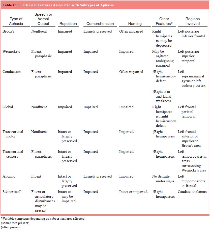
clinical and research data on aphasia. Classifications based on syndromes or
clusters of symptoms have been most favored. The major aphasia subtypes that
are now widely accepted are presented in Table 15.3. The subtypes of aphasia
have been further sub-divided in terms of their neuroanatomical loci into
perisylvian (Broca’s, Wernicke’s, conduction and global) and extrasylvian
(transcortical-motor, transcortical-sensory, mixed, anomic and subcortical)
aphasias (Benson and Geschwind, 1985). The impairment and preservation of the
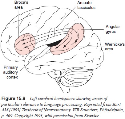
ability to repeat appear to coincide with this dichotomy. Figure 15.9 depicts
some of the regions of the left cerebral cortex that are involved in language
functioning.

Computed tomographic studies have linked aphasia
with left-sided lesions in areas lying outside the cerebral cortex. This
finding suggests that language is also subserved by subcortical areas.
Subcortical aphasia is characterized by mutism in the early stage of the
disorder, followed by articulatory disturbances and paraphasic output. Paraphasia
appears to resolve when the pa-tient has to repeat sentences. Comprehension is
commonly im-paired, and other language disturbances may also be involved. One
distinguishing feature of this type of aphasia is the transient nature of the
severe language defects seen in the early stages (Benson and Geschwind, 1985).
The neuropathology in subcorti-cal aphasia has been found to vary in terms of
localization. Most commonly, subcortical aphasia is associated with lesions in
the thalamus, caudate, and putamen (Naeser et
al., 1982).
Some inconsistent findings have raised questions
about the role of subcortical damage in aphasia. For example, it has been noted
that aphasia can resolve even in the presence of persisting subcortical
abnormalities. Conversely, in some cases, aphasia does not follow damage to
subcortical areas. In some instances, evidence from functional neuroimaging
studies has been used to understand such discrepancies. PET studies reveal that
structural damage in an area might result in hypometabolism in distant, intact
areas. Metter and coworkers (1987) found that aphasia was associ-ated with the
presence of cortical hypometabolism after structural lesions in subcortical
areas. Thus, there is some suggestion that changes in cortical hypometabolism accompanying
subcortical le-sions might be a factor associated with the presence or absence
of aphasia rather than the lesion in the subcortical region.
Related Topics