Chapter: 12th Nursing : Chapter 1 : Human Anatomy and Physiology
The Cardiovascular System
The Cardiovascular System
Heart is the master of the cardiovascular system. It pumps out
blood all over the body to circulate and supply oxygen, electrolytes, nutrients
and hormones. Blood circulation controls the body temperature, the PH or acid
base balance of the body. Circulation fights against infection, helps in
clotting of blood during injury and eliminates waste products such as carbon
dioxide.
Position
The heart lies in the thoracic cavity at the mediastinum [the space
between the lungs]. It lies obliquely a little more to the left than the right.
The apex is about 9cm to the left of the midline at the level of the 5th
intercostal space and the base extends to the level of the 2nd rib.
Organs associated with the heart
Inferiorly- The apex rest on the central tendon of the diaphragm .
Superiorly- The great vessels in the aorta, superior venacava,
pulmonary artery and pulmonary veins.
Posteriorly- Oesophagus ,trachea, left and right bronchus, decending
aorta, inferior venacava, and thoracic vertebrae.
Lateraly- The left lung overlaps the left side of the heart.
Anteriorly- The sternum, rib, and intercostal muscles.
Structure
The heart wall is composed of three layers of tissues. Namely
·
Pericardium
·
Myocardium
·
Endocardium.
Pericardium
The pericardium is the outmost layer and is made up of two sacs.
The outer sac and inner side
1. Fibrous pericardium –consists of fibrous tissue.
2. Serous pericardium- double layer of serous membrane.
The Fibrous Pericardium continues with the tunica adventitia of
the great blood vessels above and is adherent to the diaphragm. It consists of
two layers.
·
The outer layer of the serous pericardium is the parietal
pericardium
·
The inner layer is visceral pericardium adherent to the heart
muscles.
It secretes serous fluid called pericardial fluid. Pericardial
fluid present in the space between the visceral and parietal layer, which allow
smooth movement of the heart.
Myocardium
The myocardium is composed of specialized cardiac muscles found
only in the heart. The Muscle arrangement of the myocardium enable the atria
and ventricles to contract in a coordinated and efficient manner. It is also
important for electrical activity of the heart.
Endocardium
It lines chambers and valves of the heart. It is a thin, smooth
membrane to ensure smooth flow of blood through the heart. It consists of
flattened epithelial cells, and it is continuous with the endothelium lining of
blood vessels.
The cardiovascular [cardio-heart, vascular-blood vessels] system
is divided for descriptive purpose into two main parts.
·
The heart pumping action ensures constant circulation of the
blood.
·
The blood vessels which form a lengthy network through which the
blood flows.
The lymphatic system is closely connected by both structurally and
functionally with a cardiovascular system.
The heart pumps blood in to three anatomically separate systems of
blood vessels.
·
The pulmonary circulation
·
The systemic circulation
·
The coronary circulation.
Pulmonary Circulation
The portion of the circulatory system which carries deoxygenated
blood away from the right ventricle of the heart, to the lungs and returns
oxygenated blood to the left atrium and ventricle of the heart.
Systemic Circulation
Systemic circulation carries oxygenated blood from the left
ventricle, through the arteries to the capillaries in the tissues of the body.
From the tissue capillaries the deoxygenated blood returns through a system of
veins to the right atrium of the heart.
Coronary Circulation
Coronary Circulation is the circulation of blood in the blood
vessels that supplies the heart muscles.
The cardiovascular system ensures a continuous flow of blood to
all body cells. Heart plays a major role in continual physiological adjustment
to maintain an adequate blood supply.
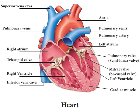
Interior of the heart
The heart is divided into a right and left side by the septum,
partition consisting of myocardium covered by endocardium. It subdivided into 4
chambers they are
·
Left atrium.
·
Right atrium
·
Left ventricle
·
Right ventricle.
Right Atrium
Right atrial chamber is located in the upper portion of the right
side of the heart which receives blood from all parts of the body by superior
and inferior venacava. They are thin less muscular walls and smaller than
ventricles.
Left Atrium
Left atrium receives oxygenated blood from the lungs and pumps it
down into the left ventricle.
The lower portion of the heart left side of is left ventricle.
Right Ventricle
Deoxygenated blood flows in the right atrium, passes through the
tricuspid valve and in to the right ventricle, which pumps the blood up to the
lungs by pulmonary artery, through the pulmonary valve.
Left Ventricle
It is larger and more muscular chamber. It pumps and delivers the
blood to all parts of the body by a larger artery [aorta] for systemic
circulation.
Flow Of Blood Through The Heart
·
The right atrium receives deoxygenated blood from all over the
body by superior and inferior venacava.
·
Deoxygenated blood from right atrium passes through right
atrioventricular valve to right ventricle.
·
Pulmonary artery collects deoxygenated blood from right ventricle
to lungs through pulmonary valve or semilunarvalve.
·
Pulmonary artery is divided in to left and right carries either
side of the lungs.
·
In lungs exchange of gases takes place.
·
Oxygenated blood passes through pulmonary veins to the left
atrium.
·
In left atrium blood passes through left atrioventricular valve to
the left ventricles carries blood from left ventricles through aortic valves.
·
Arteries-Oxygenated blood passes through all parts of the body.
Valves
These are the fibrous flaps of tissues that are present in cardiac
chamber, between the arteries chambers and veins. They ensure unidirectional
flow and prevent backflow of blood.
They are
Arterioventricular Valve
It is present in every ventricle and atrium.
Tricuspid Valve
The valve lies between the right ventricle and right atrium.
Mitral Valve
It is present between left atrium and left [bicuspid valve]
ventricle.
Semilunar Valve
It is present between arteries and ventricles.
Aortic Valve
An aortic valve is present between the aorta and left ventricle.
Pulmonary Valve
Pulmonary valve exists between the pulmonary artery and right
ventricle.
Functions of heart
The structure of the heart is primarily responsible for
transportation of blood and to supply nutrients to all parts of the body. This
continuous activity uplifts the role of the heart as a vital organ.
Blood pumping cycle of the heart is called as cardiac cycle, which
ensure that the blood is distributed throughout the body. The oxygen
distribution process begins when oxygen –free blood (impure) enters in to the
heart through the right atrium goes in to the right ventricle, enters the lungs
for oxygenation and release of carbondi-oxide and transfers in to left atrial
chambers, ready for re-distribution. About 5-6 liters of blood
circulates in the body and cardiac cycles are completed per minute.
Oxygen reloading process occur in two phase. The systole is
a short period that occurs, when the tricuspid and mitral valve close.
The diastole is a relatively longer period when the aortic
and pulmonary valve close. The systole –disastole relationship is the mean
blood pressure. Other ways of physically determining the regular functioning of
the heart is through examining the pulse rate.
Circulatory System
In the fig - 1.6 shows Red color indicates oxygenated blood
carried in arteries. Blue indicates deoxygenated blood carried in veins.
The relationship between the heart and different types of blood
vessels.
Heart
·
Aorta
·
Arteries
·
Arterioles
·
Capillaries
·
Venules
·
Veins
·
Venacava
·
Heart
Arteries
Arteries are the blood vessels that carries oxygenated blood
through out the body. Arteries consists of several layers and smooth muscles
that enable them to pump blood throughout the body after it leaves the heart.
ARTERIES - It has three layers
·
Tunica adventica
·
Tunica media
·
Tunica intima
Arterioles
Blood valve that receives blood from the arteries. Those are
present next to the arteries and before the capillaries. Arterioles also have
smooth muscles.
Capillaries
These are the smallest structure of the circulatory system. The
point at which the exchange of oxygen and corbon di-oxide occurs through the
thin walls of the capillaries.
Venules
Blood vessels that receive blood from the capillaries and
transport deoxygenated blood to the veins.
Veins
Veins are blood vessels that carry blood towards the heart. Blood
vessels receive blood from the venules and transport blood back to the heart.
Like arteries, veins, have three layers.
Blood Components
Blood is a body fluid that consists of:
·
Plasma
·
Red Blood Cells
·
White Blood Cells
·
Platelets
Plasma
Plasma in blood is over 50% of the volume of blood and over 90% of
plasma is water. The main component of plasma is plasma albumin which is a
protein that enables and controls the osmotic pressure of the blood.
Red Blood Cells (RBC)
Red blood cells or erythrocytes are disc like in shape. RBCs are
enucleated, do not contain a nucleus. The red blood cells contain iron laden
haemoglobin which carries oxygen to the cells. Red blood cells also contain
glycoproteins which determine the blood group of an individual. The blood types
are type A, type B, type AB and type O.
White Blood Cells
White blood cells or leukocytes are part of the immune system
which fights against infections from pathogens. When the white blood cell count
rises, it is a sign of infection. All leukocytes have a distinct nucleus.
The various types of white blood cells are:
·
Eosinophils
·
Basophils
·
Neutrophils
·
Lymphocytes
·
Monocytes
Platelets
Platelets or thrombocytes(do not have nucleus) maintain
haemostasis (clotting). Haemostasis is enabled by the coagulation or thickening
of blood by production of fibrin from the clotting factors found within the
platelets to prevent blood loss, when a blood vessel has been broken due to
injury.
Lymphatic system
The lymphatic subsystem, a part of the circulatory system, is
closely aligned with the body’s immune system and removes excessive fluid from
the body. The lymphatic vessels contain lymph including lymphocytes. Lymphatic
system also consists of lymph nodes and lymphatic organs. These organs include
the thymus gland, spleen, bone marrow and tonsils.
Nodes are found throughout the lymphatic system and they serve to filter the
blood as it travels throughout the body. Swollen lymph nodes are a
signal of a disease or an infection. Many lymph nodes are found
in the neck area, under the arms and in the groin area, although there are
hundreds of them throughout the body.
Spleen
Spleen is an organ present in the upper left part of the abdomen
and to the left side of the stomach. The spleen plays multiple supporting roles
in the body. It acts as a filter for blood as part of the immune system. Old
red blood cells are recycled in the spleen, platelets and white bloods are stored.
Diseases related to blood and blood vessels
·
Phlebitis
·
Deep Vein Thrombosis (DVT)
·
Anaemia, including pernicious anaemia and sickle cell anaemia
·
Leukaemia
·
Lymphoma
·
Thrombocytopenia.
Related Topics