Chapter: 11th Zoology : Chapter 9 : Locomotion and Movement
The Axial skeleton
The Axial skeleton
Axial skeleton forms the main axis of the body. It
consists of the skull, hyoid bone, vertebral column and thoracic cage.
a) The Skull
The skull is composed of two sets of bones cranial
and facial bones. It consists of 22 bones of which 8 are cranial bones and 14
are facial bones (Figure 9.7). The cranial bones form the hard protective outer
covering of the brain and called the brain box. The capacity of the cranium is
1500 cm3.. These bones are joined by sut ures which are immovable. They are a paired parietal, paired temporal and individual bones such as the frontal, sphenoid, occipital and ethmoid.
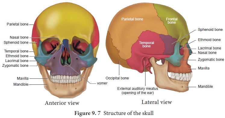
The large hole in the temporal bone is the external auditory meatus. In the facial
bones maxilla, zygomatic,
palatine, lacrimal, nasal are paired
bones whereas mandible or lower jaw and vomer are unpaired bones. They form the front
part of the skull. A single U-shaped hyoid
bone is present at the base of the buccal cavity. It is the only one bone
without any joint. Each middle ear contains three tiny bones - malleus, incus and stapes collectively
are called ear ossicles . The upper
jaw is formed of the maxilla and the
lower jaw is formed of the mandible.
The upper jaw is fused with the cranium and is immovable. The lower jaw is
connected to the cranium by muscles and is movable. The most
Foramen
magnum is a large opening found at
the posterior base of the skull. Through
this opening the medulla oblongata
of the brain descends down as the spinal cord.
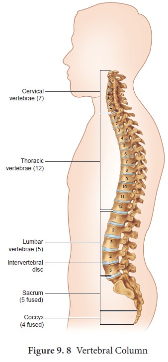
b) The Vertebral Column
Vertebral column is also called the back bone. It
consists of 33 serially arranged vertebrae which are interc onnected by
cartilage known as intervertebral disc (Figure 9.8). The vertebral column
extends from the base of the skull to the pelvis and forms the main frame work
of the trunk. The vertebral column has five major regions. They are, the Cervical, Thoracic, Lumbar, Sacrum (5 sacral vertebrae found in the
infant which are fused to form one bone in the adult) and Coccyx (4 coccygeal vertebrae found in the infant which are fused
to form one bone in the adult).
Each vertebra has a central hollow portion, the neural canal, through which the spinal cord passes. The first vertebra is called as the atlas and the second vertebra is called as the axis. Atlas is articulated with the occipital condyles.
The vertebral column protects the spinal cord,
supports the head and serves as the point of attachment for the ribs and
musculature of the back.
(c) The Sternum (Chest bone)
Sternum is a flat bone on the mid ventral line of
the thorax. It provides space for the attachment of the thoracic ribs and
abdominal muscles.
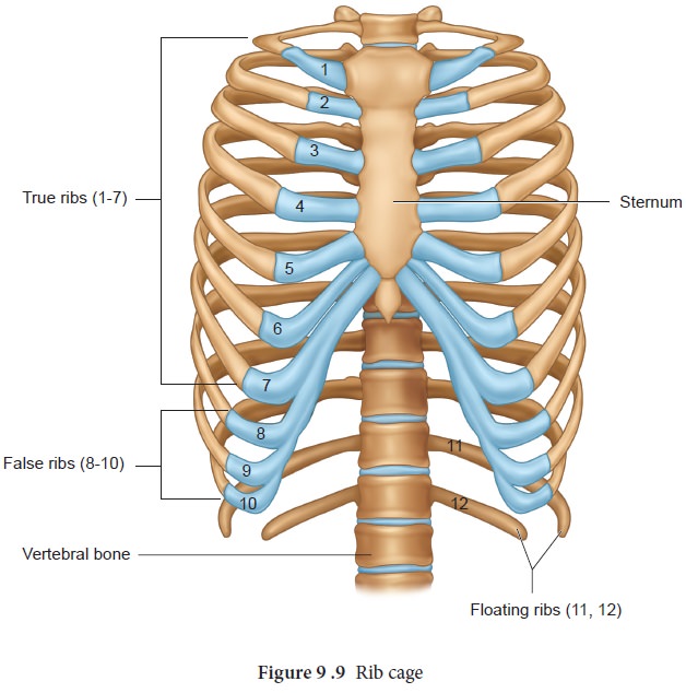
(d) The Rib cage
There are 12 pairs of ribs (Figure 9.9). Each rib is a thin flat bone connected dorsally tothe vertebral column and ventrally to the sternum. It has two articulation surfaces on its dorsal end, hence called bicephalic. The first seven pairs of ribs are called ‘true ribs’ or vertebro–sternal ribs. Dorsally they are attached to the thoracic vertebrae and ventrally connected to the sternum with the help of hyaline cartilages. The 8th, 9th and 10th pairs of ribs do not articulate directly with the sternum but joined with the cartilaginous (hyaline cartilage) part of the seventh rib. These are called ‘false ribs’ or vertebro -chondral ribs. The last 11th and 12th pairs of ribs are not connected ventrally. Therefore, they are called as ‘floating ribs’ or vertebral ribs. Thoracic vertebrae, ribs and sternum together form the ribcage.
Related Topics