Chapter: Microbiology and Immunology: Morphology and Physiology of Bacteria
Surface Appendages - Structure and Functions of Bacterial Cell Envelope
Structure and Functions of Bacterial Cell Envelope
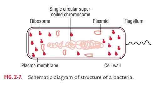
The outer layer or cell envelope provides a structural and physi-ological barrier between the protoplasm (inside) of the cell and the external environment. The cell envelope protects bacteria against osmotic lysis and gives bacteria rigidity and shape. The cell envelope primarily consists of two components: a cell wall and cytoplasmic or plasma membrane. It encloses the proto-plasm, which consists of (i) cytoplasm, (ii) cytoplasmic inclu-sions (mesosomes, ribosomes, inclusion granules, vacuoles), and (iii) a single circular DNA (Fig. 2-7).
Surface Appendages
The surface appendages of the bacteria include flagella and
fimbriae or pili.
◗
Flagella
Bacterial flagella are thread-like appendages intricately embedded
in the cell envelope. These structures are responsible for conferring motility
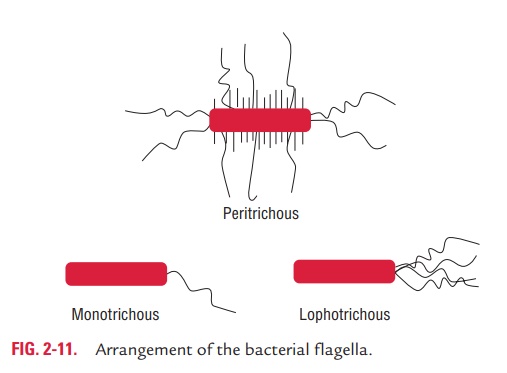
to the bacteria. The arrangement of flagella varies between different bacterial
species. Depending on the arrangement, flagella can be of the following types:
·
Monotrichous (single polar flagellum), e.g., Vibrio cholerae.
·
Lophotrichous (multiple polar flagella), e.g., Spirilla.
·
Peritrichous (flagella distributed over the entire cell), e.g., SalmonellaTyphi, E. coli, etc.
·
Amphitrichous (single flagellum at both the ends), e.g., Spirillum minus (Fig. 2-11).
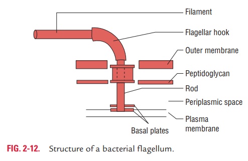
Structure: The flagella are 3–20mm in length and 0.01–0.03mmin diameter. The main part
of the filament is made up of protein subunits called flagellin arranged in
several helices around a cen-tral hollow core. The flagellum is attached to the
bacterial cell
body by a complex structure consisting of a hook and a basal body.
The basal body bears a set of rings, one pair in Gram-positive bacteria and two
pairs in Gram-negative bacteria, through which the bacteria rotates either in a
clockwise or an anticlockwise direction. Above the base of filament is the
hook, a short curved structure between the external filament and basal body.
This part produces a propeller-like repulsion from the revolving flagellum
(Fig. 2-12).
Spirochetes are motile
bacteria but without any external flagella. They are motile due to the presence
of an axial fila-ment. Axial filament consists of a bundle of flagellum-like
structures that lie between the cell surface and an outer sheath, and connects
one end of the cell to the other. They are some-times called the
endoflagellates.
Function: Flagella have the following
functions:
·
They are primarily responsible for motility of bacteria by
chemotaxis.
·
They may play a role in bacterial survival and pathogenesis.
·
They are highly antigenic, they possess H antigens, and some of the
immune responses to infection are directed against these proteins. The flagella
of different bacteria differ antigenically. Flagellar antibodies are not
protective but help in serodiagnosis.
Demonstration of flagella: The flagella can be
demonstratedby direct and indirect methods. The direct methods include direct
demonstration of capsule by electron microscope. These also include
demonstration of capsule after staining by special staining methods, such as
Ryu’s method and Hugh–Leifson’s method. Since flagella are very thin
structures, these staining methods are used to demonstrate flagella by
increasing their thickness by mordanting with tannic acid.
Indirect methods of
demonstration of flagella include demonstration of motility of the bacteria by
(a) dark-ground microscopy, (b) hanging drop method, or (c) observing
spread-ing type growth on semisolid media, such as mannitol motility medium.
◗Pili
(fimbriae)
Pili or fimbriae are synonymous for
most purposes. They are hair-like
filaments that extend from cell surface and are found almost exclusively on
Gram-negative bacteria. They are composed of structural protein subunits termed
pilins. Minor proteins termed adhesins are located at the tips of pili and are
responsible for the attachment properties.
Structure: The pili are shorter and
straighter than flagella,although the basic structure is same. Like flagella,
it consists of helics of protein called pilins,
arranged around a hollow core but without a motor. They are 0.5 mm long and 10 nm thick. They
are antigenic in nature. Pilihemagglutinate RBCs of guinea pigs and are
specifically inhibited by mannose, on the basis of which they are classified
into four types as follows:
o Type 1: These occur inE. coli, Klebsiella, Shigella,andSalmonella.They are mannose sensitive.
o Type 2: These are present inSalmonellaGallinarum andSalmonella Pullorum, devoid of any hemagglutinating oradhesive properties.
o Type 3: These are present in some
strains ofKlebsiella, Serratia,etc.
They agglutinate RBC only after heating and are man-nose resistant.
o Type 4: These are mannose resistant
and occur inProteus.
Sex pili: A specialized kind of pili
called sex pili is responsiblefor the attachment of donor and recipient cells
in bacterial conjugation. These pili are longer (10–20 mm) and vary 1–4 in number.
The sex pili are of two types:
o
F pili: They specifically adsorb male specific RNA and
DNAbacteriophages. They are encoded by sex factor F and fertil-ity
inhibition–positive resistance factors (fi 1 R factors).
o
I pili: They adsorb male specific filamentous DNA
phages,encoded by col factor and fi 2 R factor.
Function: Pili play a major role in the
adherence of symbioticand pathogenic bacteria to host cells, which is a
necessary step in initiation of infection. Transfer of bacterial DNA takes
place through sex pili during the process of conjugation.
Demonstration of pili: The pili can be detected:
o Directly by electron
microscope and
o By agglutination of RBCs of
guinea pigs, fowl, horses, and pigs. They agglutinate human and sheep RBCs
weakly. The hemagglutination can be specifically inhibited by D-mannose.
Some of the Gram-positive bacteria do not possess typical pili but
instead possess a fine fibrillar arrangement of proteins on their surfaces
known as fibrils. These fibrils bind to the host surfaces. M-protein of S. pyogenes is an example of
Gram-positive bacteria possessing fibrils.
Related Topics