Cell Biology - Subcellular organelles | 11th Biochemistry : Chapter 1 : Basic Concepts of Bio Chemistry and Cell Biology
Chapter: 11th Biochemistry : Chapter 1 : Basic Concepts of Bio Chemistry and Cell Biology
Subcellular organelles
Subcellular organelles
An
eukaryotic cell does not have a homogeneous internal environment but is divided
into two major compartments ,cytoplasm and nucleus and subsequently into
individual compartments, each of which is surrounded by a membrane, addressed
as organelles.
1. Cell Membrane
All
plants, animal cells, prokaryotic cells, and fungal cells are bounded by a cell
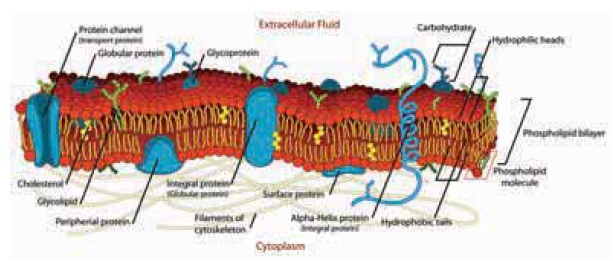
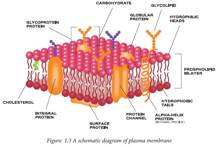
membrane, which is sometimes known as plasma membrane.
Chemical
composition of Cell membrane: Cellular membranes including plasma membranes and
internal membranes consist of mainly lipid, protein and water. Lipids
constitute about 40 percent of the membrane composition. Lipids are complex
mixtures of cholesterol and fatty acid esters, mainly in the form of glycerides
and phospholipids. Glycerol is a three-carbon molecule that forms the backbone
of membrane lipids. Within an individual glycerophospholipid, fatty acids are
attached to the first and second carbons, and the phosphate group is attached
to the third carbon of the glycerol backbone.
Lipid
bilayer encircles a cell and is amphipathic with one end as a hydrophilic
‘head’ and the other end as a hydrophobic ‘tail’. Each ‘leaf ’ of the lipid
bilayer has one side consisting of an array of the hydrophilic heads, while the
other side consists of the hydrophobic tails. An aqueous environment causes the
hydrophobic tails to aggregate, so that the hydrophobic sides of each leaf come
together to form a non-ionic centre, like an oil drop in water. The hydrophilic
end of the two leaves face into the ionic milieu on either side of the lipid
bilayer. The lipid bilayer has the important property of fluidity which allows
it to fuse with other membranes, generate new membranes by fission, and provide
solvent for proteins that can reside within the layer and move around within
it. It can permit water but will not permit ions, small charged molecules, and
all large molecules.
The
plasma membrane separates the contents of the cell from the external
environment. In the unicellular organism, the ‘external environment’ is the
exterior world; for a multicellular organism, it is both the exterior world
outside the organism as well as the interior world created by other cells. For
this process of division of a pre-existing cell, a cell must carry within it
the information for reproducing all its components. The form of this
information is a single type of genetic material, DNA, which codes for all the
proteins of the cell.
Functions of cell membrane
· It serves to keep all the component parts of the
cell together in one place.
· It regulates the continuous movement of substances
into and out of the cell.
·
It can serve as a
base of attachment for the cytoskeleton in some organisms and the cell wall in
others. Thus, the cell membrane also serves to maintain its shape.
·
It can regulate the
cell growth through the balance of endocytosis and exocytosis.
·
It can maintain the
concentration of water, inorganic ions and organic molecules between the cell
and the environment.
·
The plasma membrane
also receives signals and coordinates molecular interactions at the surface
such as cell to cell recognition, adhesion and communication.
2. Cell wall
The cell wall is a non-living rigid
structure that forms an outer covering for the plasma membrane of fungi and
plants. Cell wall not only gives shape to the cell and protects the cell from
mechanical damage and infection, it also helps in cell-to-cell interaction and
provides a barrier to undesirable macromolecules.
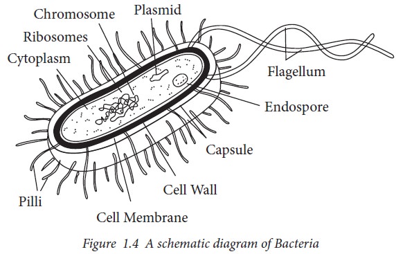
Bacterial cell wall:
Bacteria
have a cell wall, which is a rigid, carbohydrate-containing structure that
surrounds the bacterial cell. However, the genus Mycoplasma, do not have cell
wall. The cell wall provides the bacteria with several benefits including
protection of the bacterium from damage by encircling it with a tough, rigid
structure. This structure is also porous. Small molecules are able to freely
pass through the cell wall to the membrane, but large molecules are excluded.
By performing this function, the cell wall acts as a coarse filter. The primary
function of the cell wall, however, is to maintain the cell shape and prevent
bursting due to osmotic pressure (called lysis).
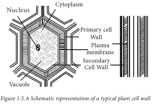
Plant cell wall
Algae
have cell wall, made of cellulose, galactans, mannans and minerals like calcium
carbonate, while in other plants it consists of cellulose, hemicellulose,
pectins and proteins. The cell wall of a young plant cell, the primary wall is
capable of growth, which gradually diminishes as the cell matures and the
secondary wall is formed on the inner (towards membrane) side of the cell. The
middle lamella is a layer mainly of calcium pectate which holds or glues the
different neighbouring cells together. The cell wall and middle lamellae may be
traversed by plasmodesmata which connect the cytoplasm of neighbouring cells.
The main functions of the cell wall are:
· Cell wall provides structural and mechanical
support.
· Cell wall determines and maintains the shape of the
plant cell and governs plant architecture
· Cell wall
resists internal turgor pressure of cell.
· Cell wall
regulates growth rate and diffusion of materials.
· Cell wall
functions as stores of carbohydrates.
· Cell wall
protects against pathogens, dehydration, and other environmental factors.
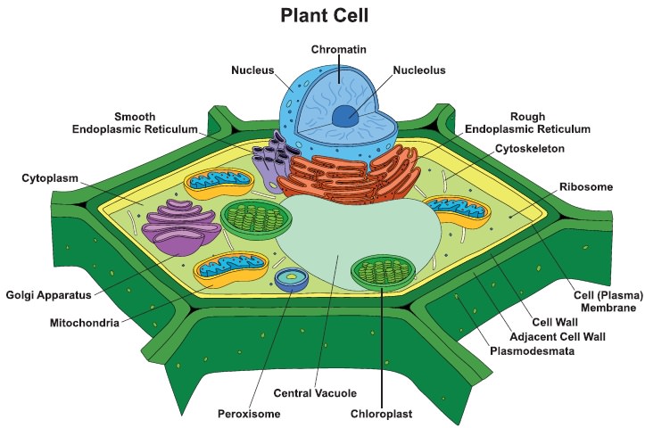
3. Nucleus
The
largest organelle in the cell is nucleus which is enveloped by bound double
layered nuclear membrane preserving the genetic material called chromatin.
Nucleus occupies 1%-2% and 10% in yeast and animal cells respectively. The
genetic material forms a mass called chromatin that is concentrated in one part
of the nucleus. The outer and inner membranes are separated by lumen. The outer
membrane of the nuclear envelope is continuous with the endoplasmice reticulum
(ER) membrane, and the lumen of the nuclear envelope is continuous with the
lumen of the ER. The inner nuclear membrane is usually supported by a network
of filaments called the nuclear lamina, located in the nucleus and anchored to
the inner membrane. The nucleus contains subcompartments with specialized
functions and the major subcompartment in the nucleus is the nucleolus.
The pores of nuclear membranes are
large enough to be completely permeable to smaller molecules, so there is no
difference in the aqueous environment of the nucleus and the cytoplasm. The
nucleus is considered to be the core of the cell which regulates all metabolic
events.
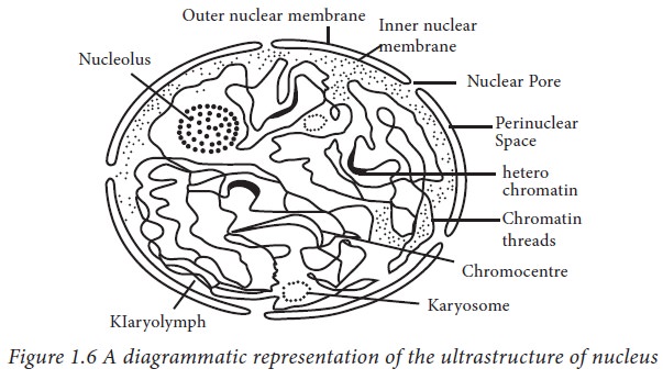
Nuclear envelope: The nucleus is separated from the cytoplasm by a double membrane, the nuclear envelope and
the two membranes separated from each other by a perinuclear space of varying
width. There are little holes in the nuclear envelope called nuclear pores
which help the substances to move into or out of the nucleus. DNA occupies most
of the space inside a nucleus. DNA is the genetic material and provides the
instructions essential for building proteins. Proteins are responsible for
helping with most activities in a cell. Inside the nucleus is a round body
called nucleolus, which is present in a eukaryotic cell. The nucleolus is
devoid of an encircling membrane. The nucleolus produces the ribosomal subunits
from proteins and ribosomal RNA, also known as rRNA. It then sends the subunits
out to the rest of the cell where they combine into complete ribosomes.
Ribosomes make proteins; therefore, the nucleolus plays a vital role in making
proteins in the cell.
4. Mitochondria- the power houses of the cell
A cell has a compartment for energy
production. It obtains energy from the food supplied by its environment. This
energy then has to be converted into some form that can be distributed
throughout the cell. The common solution is to store energy in the form of a
common molecule that can be used whenever and wherever it is needed in the
cell. The term ‘mitochondrion’ is derived from the Greek word ‘mitos’ which
means ‘thread’ and ‘chondrion’ which means ‘granule’. Mitochondria is a
membrane bound cellular structure and is found in most of the eukaryotic
aerobic cells. Mitochondria may assume different shapes ranging from granular
to filamentous depending upon the functional state of the cell. They are
spherical in yeast cells, elliptical in kidney cells, elongated in liver cells
and filamentous in fibroblasts. The size of the mitochondria ranges from 0.5 to
1.0 μm in diameter.
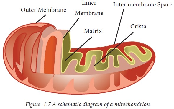
The mitochondria consist of a smooth
outer membrane, which has a large number of special proteins known as the
porins, separated by a space from an inner membrane. The inner membrane is
thrown into folds or invagination called cristae which extend into matrix, the
mitochondrial lumen. Both the membranes are separated by a clear inter membrane
space. The cristae are irregularly shaped like villous and finger like
projections. The membranes are made up of phospholipids and proteins.
Functions of Mitochondria
· The mitochondria can help the living cell to
convert energy supplied by the environment into ATP, the common molecule,
required for chemical reactions. ATP can be generated in two pathways: in the
cytosol, and in mitochondrion. First pathway exists in the cytosol of an
eukaryotic cell (or within a bacterial cell) where glycolysis degrades glucose
to lactate and releases two molecules of ATP.
· Second pathway is the main source of energy
production as ATP (called oxidative phosphorylation and involves the electron
transport chain). Pyruvate generated from glycolysis enters the matrix (lumen)
of the mitochondrion, where it is degraded and combined with coenzyme A to form
acetyl CoA. The acetyl part of the acetyl CoA is then degraded to carbon
dioxide by the citric acid cycle, releasing hydrogen atoms. The hydrogen atoms
are used to reduce the carrier NAD+ to NADH, and then oxidation of
NADH releases a proton and an electron.
· Mitochondria
help the cells
to maintain proper concentration of calcium ions within the compartments
of the cell.
· Mitochondria also help in erythropoiesis and
biosynthesis of hormones like testosterone and estrogen.
· The mitochondria of liver cells have enzymes that
detoxify ammonia.
· The mitochondria also play an important role in the
process of apoptosis or programmed cell death. Abnormal death of cells due to
the dysfunction of mitochondria can affect the function of an organ.
· The mitochondria are involved in
other cellular activities
like signalling, cellular differentiation and cell senescence. They
also regulate the control of cell cycle and cell growth.
· Unlike the outer membrane, the inner membrane is
strictly permeable, it is permeable only to oxygen, ATP and it also helps in
regulating transfer of metabolites across the membrane.
· The matrix of the mitochondria is a complex mixture
of proteins and enzymes. These enzymes are important for the synthesis of ATP
molecules, mitochondrial ribosomes, tRNAs and mitochondrial DNA.
· Mitochondria also affect human health. Mitochondrial disorders
and cardiac dysfunction also play an important role in the aging process.
5. Endoplasmic reticulum (ER):
Eukaryotic
cells contain several interrelated membrane-bound compartments, collectively
termed as ‘endomembrane system’ or ER.It is a continuous membrane, which is
present in both plant cells, animal cells and absent in prokaryotic cells.There
is a series of convoluted membrane sheets which are contiguous with the outer
membrane of the nuclear envelope. This series of membrane delimited
compartments in a typical eukaryotic cell are related and interact with one
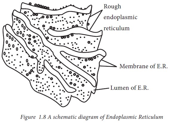
another by fission and fusion of their membranes. The space, which is present
in the endoplasmic reticulum, is called as the lumen.
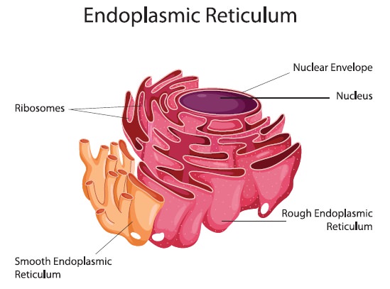
There are three morphological patterns in ER.
a. Granular or Rough endoplasmic reticulum
b. Smooth endoplasmic Reticulum
c.
Lamellar and
Vesicular endoplasmic reticulum
The
rough endoplasmic reticulum contains ribosome attached to the cytoplasmic side
of the membrane and it forms a lace like system. The smooth Endoplasmic
reticulum lacks the attached ribosome and it forms tubular structures.
The major functions of Endoplasmic reticulum are:
• They
play a vital role in the formation of the skeletal framework
• They
provide the increased surface area for cellular reactions
• They
help in the formation of nuclear membrane during cell division
• They play a vital role in the synthesis of proteins, lipids, glycogen and other steroids like cholesterol, progesterone, testosterone, etc.
• They are responsible for the secretion, synthesis, modification
and transportation of proteins and other carbohydrates to another organelle,
which includes lysosomes, Golgi apparatus, plasma membrane, etc.
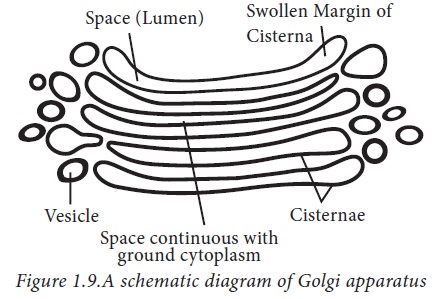
6. Golgi Apparatus
Camillo
Golgi (1898) had made the first report on the densely stained reticular
structures near the nucleus. Hence these were later named Golgi bodies,
attributed to him. They consist of many flat, disc-shaped sacs or cisternae of
0.5μm to 1.0μm diameter. These are stacked parallel to each other. Varied
numbers of cisternae are present in a Golgi complex. The Golgi cisternae are
concentrically arranged near the nucleus with distinct convex cis or the forming face and concave trans or the maturing face.
The cis and the trans faces
of the organelle are entirely different, but interconnected. The golgi
apparatus principally performs the function of packaging materials, to be
delivered either to the intra-cellular targets or secreted outside the cell.
Materials to be packaged in the form of vesicles from the ER fuse with the cis face of the golgi apparatus and move
towards the maturing face. This explains, why the golgi apparatus remains in
close association with the endoplasmic reticulum. A number of proteins
synthesized by ribosomes on the endoplasmic reticulum are modified in the
cisternae of the golgi apparatus before they are released from its trans face. Golgi apparatus is the
important site of formation of glycoproteins and glycolipids.
Functions of Golgi apparatus
a. Golgi apparatus helps in protein sorting from one compartment to
another by the secretory pathway.
b. Covalent modifications of proteins involving the addition of
small sugar molecules occur in the ER and Golgi apparatus.
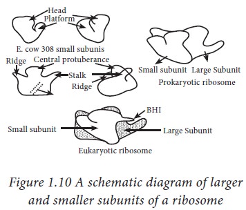
7. Ribosomes:
Ribosomes are the granular structures
first observed under the electron microscope as dense particles by George
Palade (1953). In the word ribosome, the pharse ‘ribo’ is derived from
ribonucleic acid and ‘somes’ from the Greek word ‘soma’ which means ‘body’.
Ribosomes are tiny particles about 200 Å. They are composed of ribonucleic acid
(RNA) and proteins. Ribosomes are not considered as organelles because of the
lack of a membrane around them. However, when they are producing certain proteins
they can become bound to the endoplasmic reticulum membrane. Free floating
ribosomes are also present. Ribosomes are composed of both RNA and proteins.
About 37 - 62% of ribosomes are made up of RNA and the rest is proteins. There
are two types of ribosomes based on their sedimentation properties. Prokaryotes
possess 70 S ribosomes and Eukaryotes possess 80 S ribosomes. The subunits of
ribosomes are named owning to their sedimentation rate measured as special
Svedberg Unit (‘S’). The ribosomes share a core structure which is similar to
all ribosomes despite differences in their size. The ribosomes are made up of
two subunits - a small and a large subunit. The small subunit reads the mRNA
while the large subunit joins the amino acids to form a chain of polypeptides.
Functions of ribosomes:
· The bound and the free ribosomes are similar in structure and
are involved in protein synthesis.
·
The location of the
ribosomes in a cell is a determining factor of the type of protein produced. If
the ribosomes are free floating throughout the cell, the proteins that are used
within the cell are produced. When ribosomes are attached to endoplasmic
reticulum (referred as rough endoplasmic reticulum or rough ER), the proteins
that are used inside the cell or outside the cell are produced.
·
The catalytic
activity of the ribosome is carried out by the RNA.
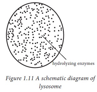
8. Lysosomes
These are membrane bound vesicular
structures formed by the process of packaging in the golgi apparatus. The
isolated lysosomal vesicles have been found to be very rich in hydrolytic
enzymes, called hydrolases such as lipases, proteases, carbohydrases, which are
optimally active at the acidic pH. These enzymes are capable of digesting
carbohydrates, proteins, lipids and nucleic acids.
9. Peroxisome:
Peroxisomes are microbodies that are
abundantly present in mammalian liver and kidney, and also in plant cells. It
depends on the type of eukaryotic cell. The matrix of Peroxisomes is rich in
enzymes but a few enzymes are located in the membrane. The common enzymes
present in the matrix of peroxisomes are catalases and peroxidases which
metabolize a number of substrates. Enzymes present in the membrane of
peroxisomes are cytochrome b5 and NADH cytochrome b5 reductase.
Functions of peroxisomes
· A major function of the peroxisome, in yeast and
plant cells are to breakdown the fatty
acid molecules, in a process called beta-oxidation. Peroxisomes are involved in
lipid biosynthesis
· Peroxisomes contain enzymes required for the
synthesis of plasmalogens
·
Peroxisomes in
seeds are responsible for the conversion of stored fatty acids to
carbohydrates, which is critical in providing energy and raw materials for
growth of the germinating plant.
10. Cytoplasm:
The
ground substance that fills the interior of the cell is called cytosol or
cytoplasm. It is a jelly-like substance and it is made up of eighty percent
water and is usually clear. It appears as a transparent and colourless fluid.
The cytoplasm serves as a molecular soup. It is in the cytoplasm where all the
cellular organelles are suspended and are bound together by a lipid bilayer
membrane. The cytoskeleton present in the cytoplasm gives the cell its shape.
Cytoplasm also constitutes numerous salts and is a very good conductor of electricity.
Various metabolic activities occur in
the cytoplasm. Metabolic pathways like glycolysis and cellular processes like
cell division take place in the cytoplasm.
·
Cytoplasm shows
differential staining properties, the areas stained with the basic dyes are the
basophilic areas of the cytoplasm and are termed as ergatoplasm for this
material.
· It is a heterogenous mixture of opaque granules and
organic compounds which gives it its colloidal nature.
· The cytoplasm conatains dissolved nutrients and it
aids to dissolve waste products.
· It helps movement of the cellular materials around
the cell through a process called cytoplasmic streaming.
· The peripheral zone of cytoplasm is jelly-like and
is known as the plasmogel. The surrounding area of the nuclear zone is thin and
liquefied in nature and is known as the plasmosol.
· The physical nature of cytoplasm is colloidal. It
has a high percentage of water and particles of various shapes and sizes are
suspended in it.
· It also contains proteins, of which 20-25 percent
are soluble proteins including enzymes.
· Also, certain amount of carbohydrates, RNAs,
inorganic salts and lipid substances are found.
· The plasmogel part of the cytoplasm is capable of
absorbing water and removing it, according to the cell's need.
·
The stomatal guard
cells present in the leaves exhibit this property.
·
An organized system
of fibres can be observed by specific staining techniques.
11. Plastids
Plastids are found in all plant cells
and in euglenoides. These are easily observed under the microscope as they are
large. They bear some specific pigments, thus imparting specific colours to the
plants. Based on the type of pigments
plastids can be classified into
different types:
Protoplastids, Amyloplastids,
Leucoplastids, Etioplasts, Chloro-amyloplasts and Chromoplasts.
• Protoplasts
contain brown carotenoids, chlorophyll a and chlorophyll c pigments
• Amyloplasts
synthesizes starch and stores them as granules in the stroma. Some types of
plastids contain enzymes for the synthesis of certain small compounds.
• The
leucoplasts are the colourless plastids of varied shapes and sizes.
• Rhodoplasts
contain chlorophyll a and chlorophyll d along with phycobilin and phycoerythrin
pigments.
• Chloroplasts-occur
in green plants are characterised by the presence of Chlorophyll a and
Chlorophyll b.
• Chromoplasts
synthesize and store pigments called carotenoids, which are red, orange, or
yellow molecules that give some flowers and fruits their colour.
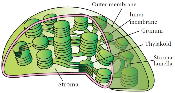
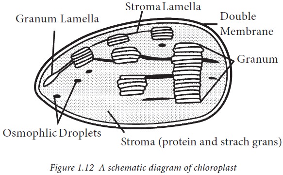
12. Chloroplasts
Chloroplasts
are members of a group of plant organelles collectively known as plastids.
These are associated with photosynthesis. Majority of the chloroplasts of the
green plants are found in the mesophyll cells of the leaves. These are
lens-shaped, oval, spherical, discoid or even ribbon-like organelles having
variable length (5-10 jm) and width (2-4 μm). Their number varies from 1
per cell of the Chlamydomonas, a
green alga to 20-40 per cell in the mesophyll. Of the two, the inner
chloroplast membrane is relatively less permeable. The space limited by the
inner membrane of the chloroplast is called the stroma. A number of organised
flattened membranous sacs called the thylakoids, are present in the stroma.
Thylakoids are arranged in stacks like the piles of coins called grana
(singular: granum) or the intergranal thylakoids. In addition, there are flat
membranous tubules called the stroma lamellae connecting the thylakoids of the
different grana. The membrane of the thylakoids enclose a space called a lumen.
The stroma of the chloroplast contains enzymes required for the synthesis of
carbohydrates and proteins. It also contains small, double-stranded circular
DNA molecules and ribosomes. Chlorophyll pigments are present in the
thylakoids.
The
thylakoids in chloroplasts contain chlorophyll and carotenoid pigments which
are responsible for trapping light energy essential for photosynthesis.
Chloroplasts develop in the parts of a plant, such as leaves, in which light
gathering and photosynthesis will occur. Plants that are grown in the dark do
not develop chloroplasts but instead develop a different type of plastid in
their leaves. Chloroplasts develop into chromoplasts when tomatoes ripen from
green to red and when green leaves of deciduous trees turn redorange or yellow.
Functions of chloroplast
· Chloroplasts function as the food producers of the
cell and every green plant in the planet is working to convert the solar energy
into sugars.
· They are responsible for breaking down the
nutrients and sugars that the cell receives and convert that into energy.
· It enables a plant to make ATP from a system in
which the electrons are provided by chlorophyll that have been activated by
light.
13. Vacuole:
The
vacuole is the membrane-bound space found in the cytoplasm. Plant cells possess
a well-developed vacuolar system, which becomes more prominent in maturing
cells. It is also present in the cells of animals, fungi and bacteria but they
are smaller in size. In plant cells the vacuoles can occupy up to 90 percent of
the volume of the cell. Vacuoles contain water, sap, excretory product and
other materials not useful for the cell. The vacuole is bound by a single
membrane called tonoplast. In plants, the tonoplast facilitates the transport
of a number of ions and other materials against concentration gradients into
the vacuole, hence, their concentration is significantly higher in the vacuole
than in the cytoplasm. In Amoeba, the contractile vacuole is important for
excretion. In many cells, as in protists, food vacuoles are formed by engulfing
the food particles.
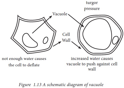
In plant
cells, the vacuoles accumulate a high concentration of sugars and other soluble
compounds. Water enters the vacuole to dilute these sugars, generating
hydrostatic pressure that is counterbalanced by the rigid wall. In this way the
cells of the plant become stiff or turgid, in the same way that when an inner
tube is inflated inside a bicycle tyre the combination becomes stiff. Vacuoles
are generally pigmented. The beautiful colors of petals and fruits are due to
presence of compounds such as the purple anthocyanins in the vacuole.
Functions of vacuole:
· Vacuoles aid in storing salts, nutrients, pigments,
minerals, proteins, facilitating the growth of the plant and playing a vital
structural role for the plant.
· It serves in other functions such as protection,
storage organelles for metabolites, growth and disposal of toxic excretory
substances.
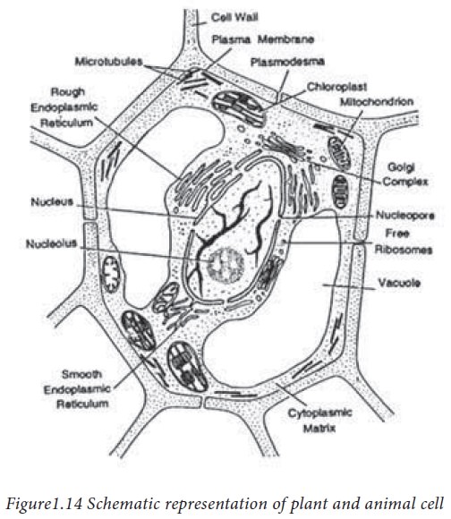
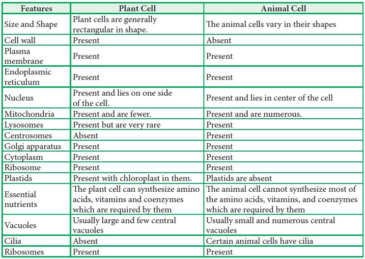
14. Distinguishing features of Plant and Animal Cells
Related Topics